All talks & lectures

Conference ·

Using MRI from Clinical Practice to Inform Prognosis

A MSToronto 2026 talk on routine MRI, AI-derived biomarkers and the evidence needed to inform prognosis in multiple sclerosis.

MSToronto 2026 · Joint ACTRIMS–ECTRIMS Meeting · Toronto, Canada
Opening slide for “Using MRI from Clinical Practice to Inform Prognosis” at MSToronto 2026 · Joint ACTRIMS–ECTRIMS Meeting
Opening slide from the original presentation
Delivered
Event
MSToronto 2026 · Joint ACTRIMS–ECTRIMS Meeting
Place
Toronto, Canada
Format
Conference
Published deck
17 slides

From the slides

Deck notes

Presented by Arman Eshaghi at MSToronto 2026, this talk follows MRI from diagnosis and monitoring to prognosis, then considers what routine hospital data and AI could contribute to a learning health system.

Deck outline

  • Prognosis, injury and reserve
  • MRI measures and the evidence for disability prediction
  • AI lesion detection and biomarkers from routine scans
  • Data-driven subtypes and stages
  • Patient benefit, fairness and clinical readiness
  • Learning from routine care and the MS-PINPOINT federated network
  • Multimodal models, take-home messages and acknowledgements

Original deck

Slide by slide

Read the 17-slide presentation in order, or use the index to jump.

  1. Slide 01

    Using MRI from Clinical Practice to Inform Prognosis
    Slide 1: Using MRI from Clinical Practice to Inform PrognosisOpen full-size slide

    Artificial Intelligence Applications from Diagnosis to Prognosis

    Arman Eshaghi, MD, PhD

    NIHR Advanced Fellow

    Queen Square Institute of Neurology, UK

  2. Slide 02

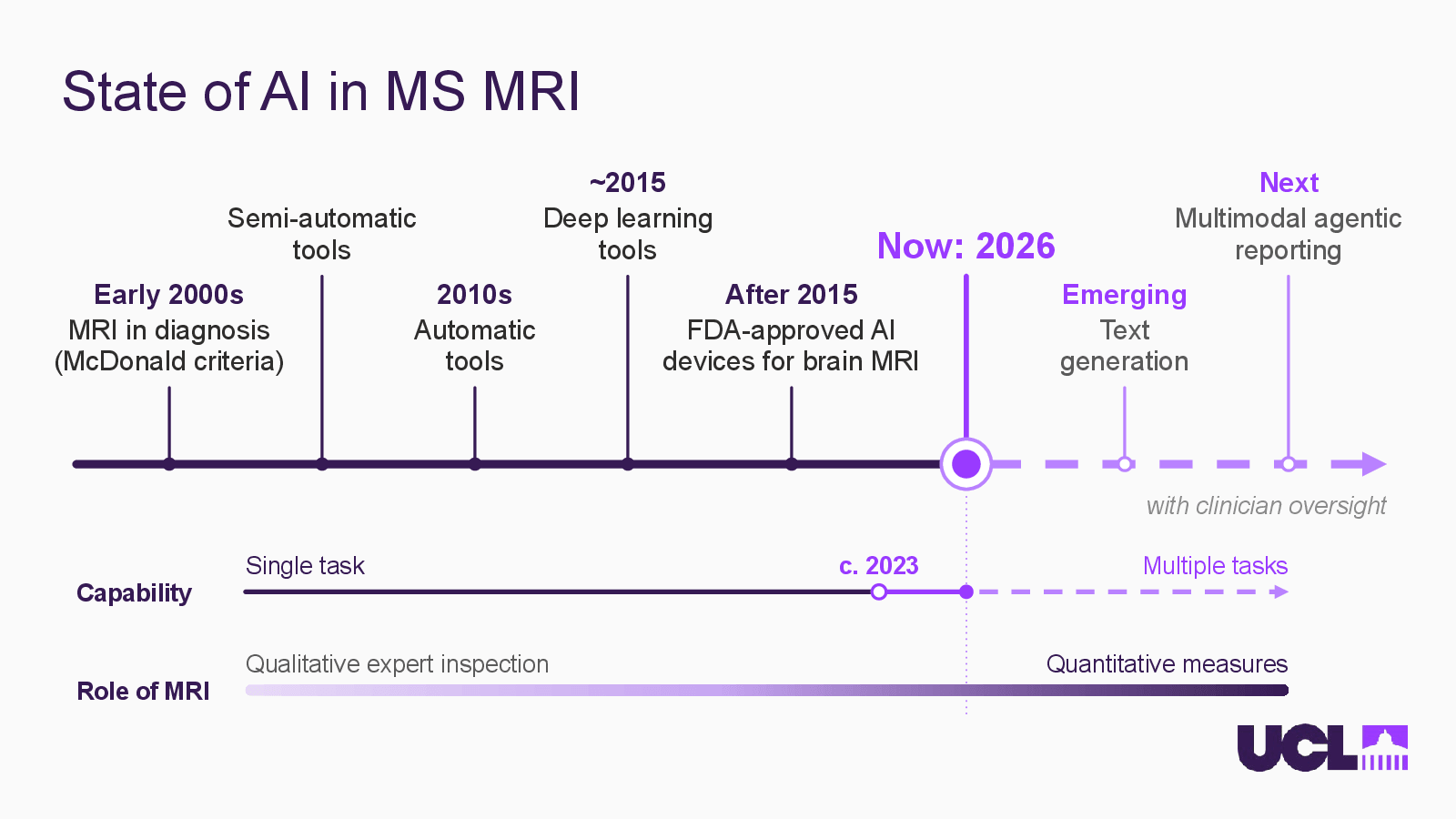
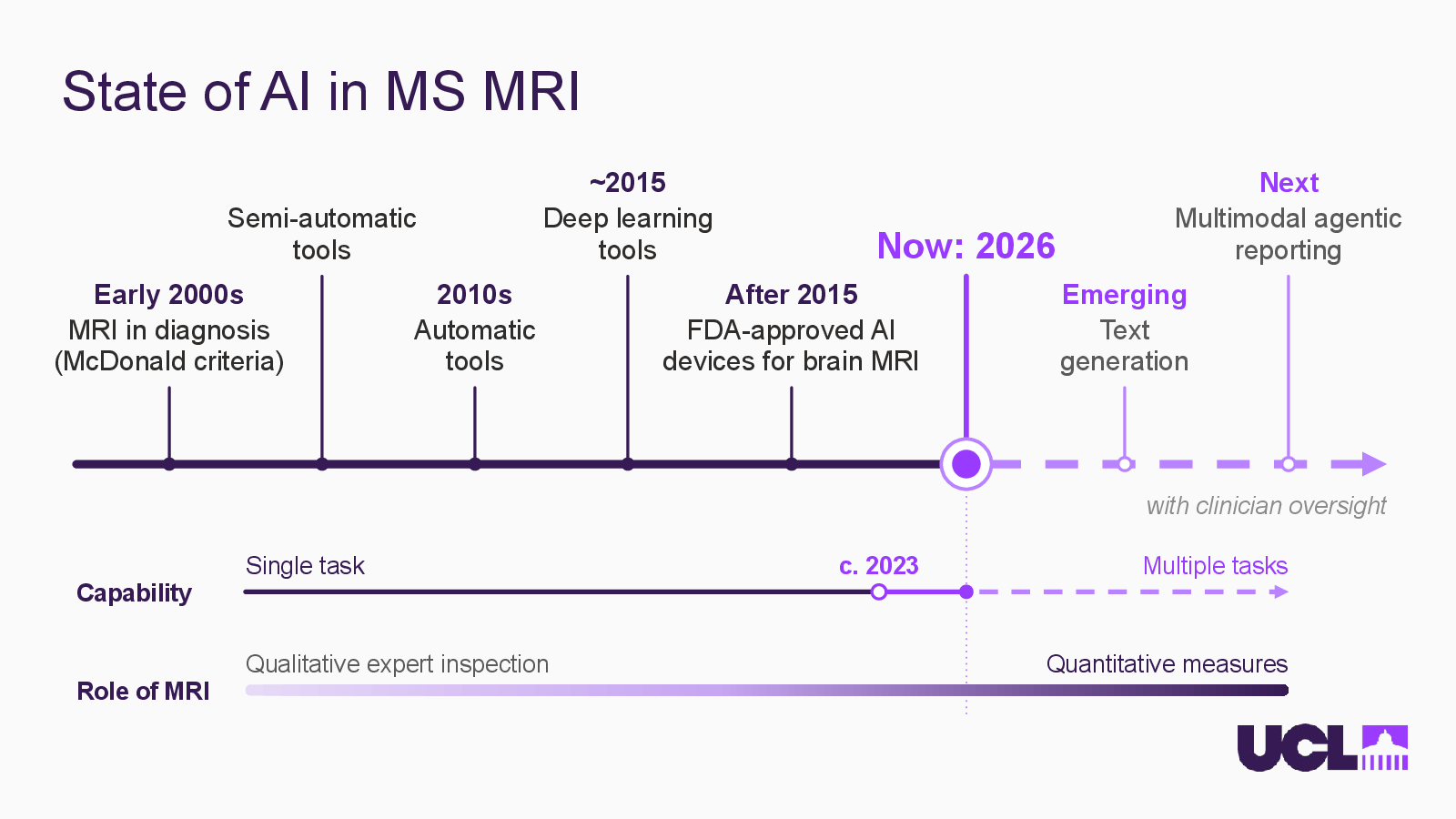
    State of AI in MS MRI

    Slide 2: State of AI in MS MRIOpen full-size slide

    Semi-automatic

    tools

    ~2015

    Deep learning

    Next

    Multimodal agentic

    reporting

    Now: 2026

    Early 2000s

    MRI in diagnosis

    (McDonald criteria)

    2010s

    Automatic

    After 2015

    FDA-approved AI

    devices for brain MRI

    Emerging

    Text

    generation

    with clinician oversight

    Single task

    c. 2023

    Multiple tasks

    Capability

    Qualitative expert inspection

    Quantitative measures

    Role of MRI

  3. Slide 03

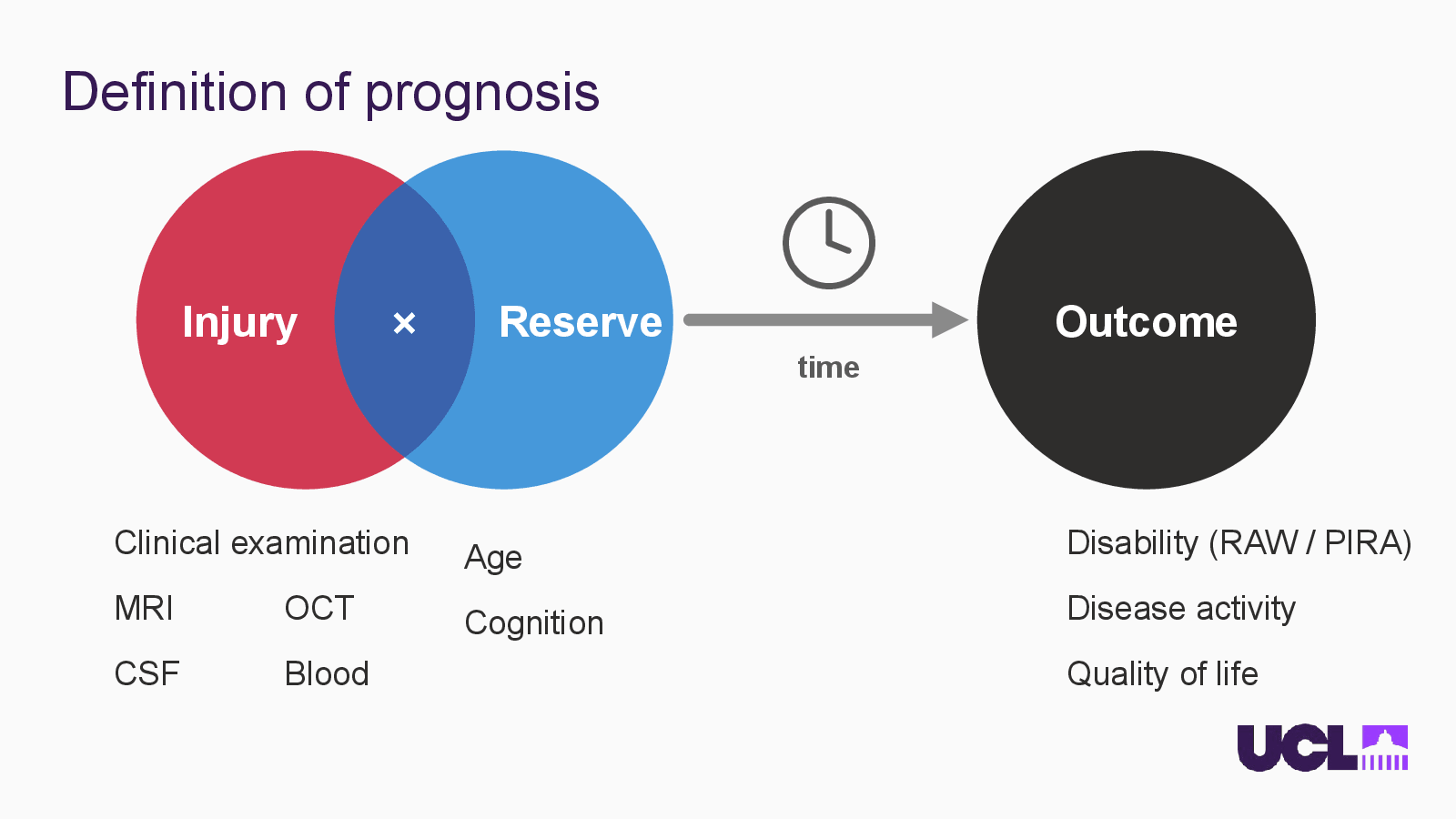
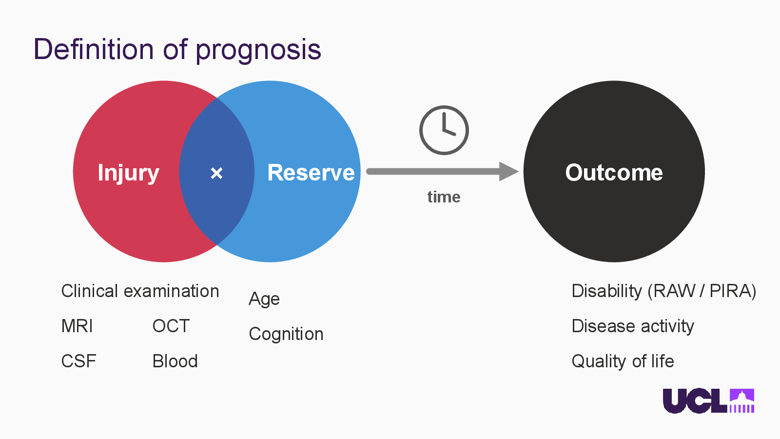
    Definition of prognosis

    Slide 3: Definition of prognosisOpen full-size slide

    Injury

    ×

    Reserve

    Outcome

    time

    Clinical examination

    Disability (RAW / PIRA)

    Age

    MRI

    OCT

    Disease activity

    Cognition

    CSF

    Blood

    Quality of life

  4. Slide 04

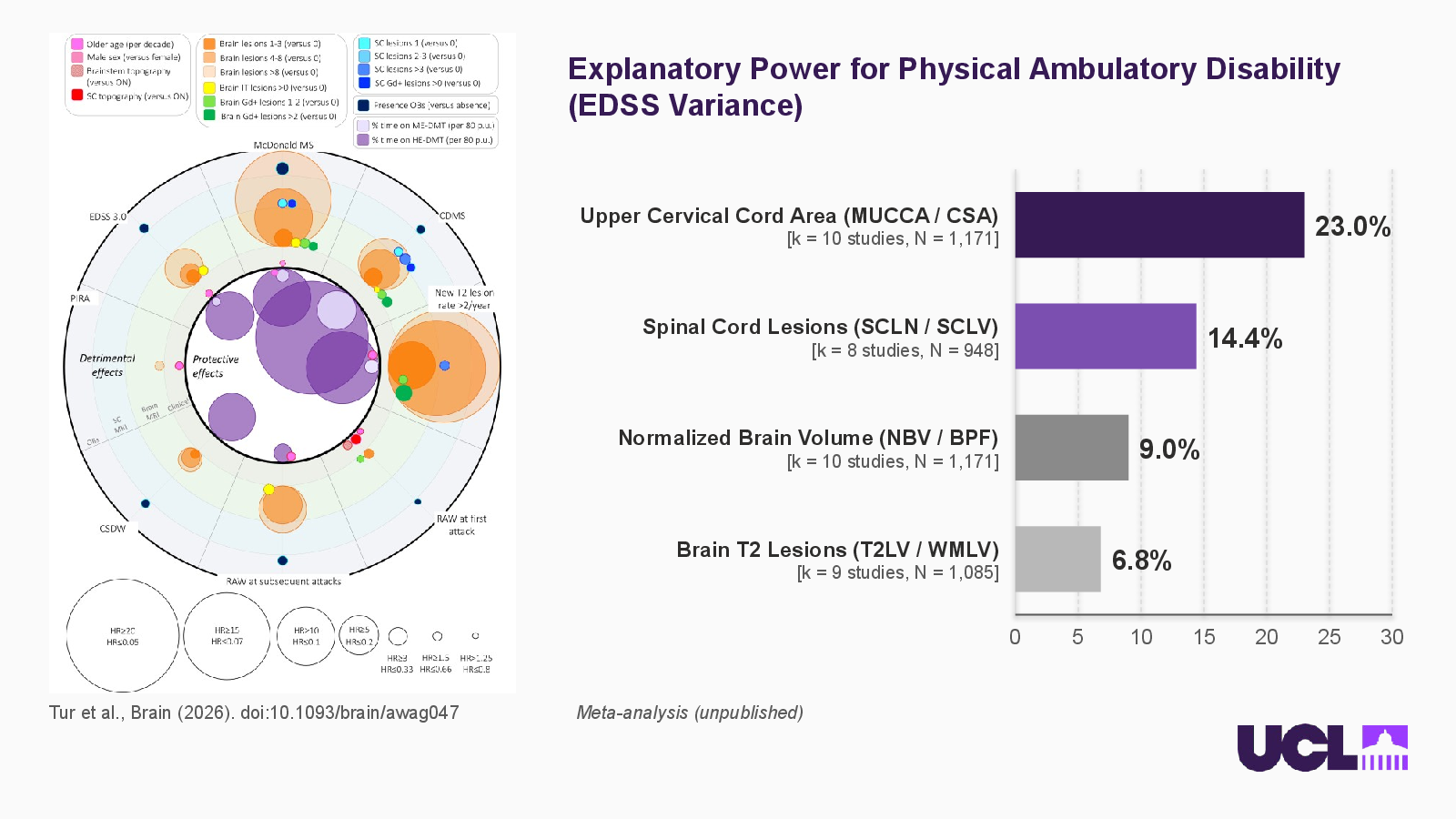
    Explanatory Power for Physical Ambulatory Disability (EDSS Variance)
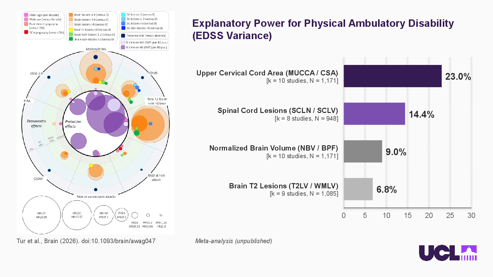
    Slide 4: Explanatory Power for Physical Ambulatory Disability (EDSS Variance)Open full-size slide

    Tur et al., Brain (2026). doi:10.1093/brain/awag047

    Explanatory Power for Physical Ambulatory Disability (EDSS Variance)

    Upper Cervical Cord Area (MUCCA / CSA)

    [k = 10 studies, N = 1,171]

    23.0%

    Spinal Cord Lesions (SCLN / SCLV)

    [k = 8 studies, N = 948]

    14.4%

    Normalized Brain Volume (NBV / BPF)

    9.0%

    Brain T2 Lesions (T2LV / WMLV)

    [k = 9 studies, N = 1,085]

    6.8%

    Meta-analysis (unpublished)

    References

  5. Slide 05

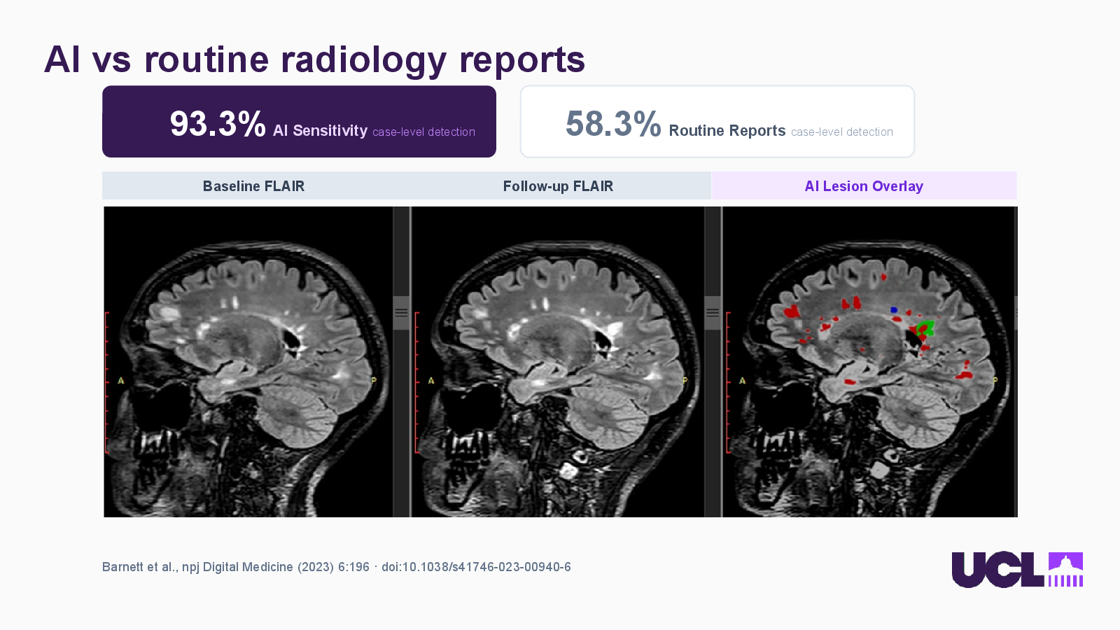
    AI vs routine radiology reports
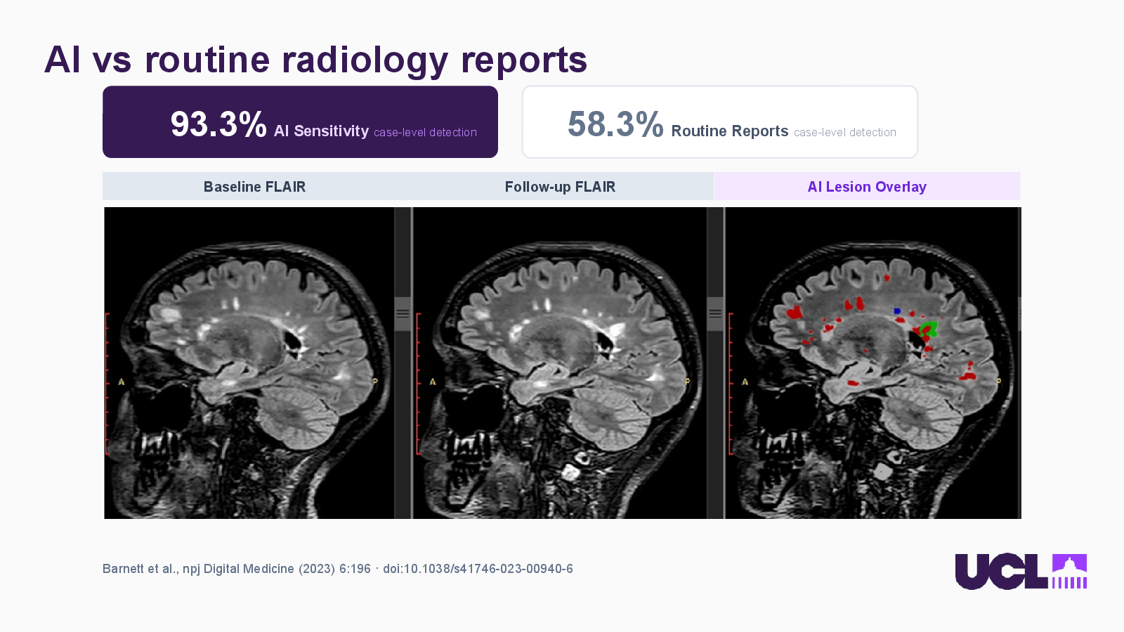
    Slide 5: AI vs routine radiology reportsOpen full-size slide

    93.3% AI Sensitivity case-level detection

    58.3% Routine Reports case-level detection

    Baseline FLAIR

    Follow-up FLAIR

    AI Lesion Overlay

    Barnett et al., npj Digital Medicine (2023) 6:196 · doi:10.1038/s41746-023-00940-6

    References

  6. Slide 06

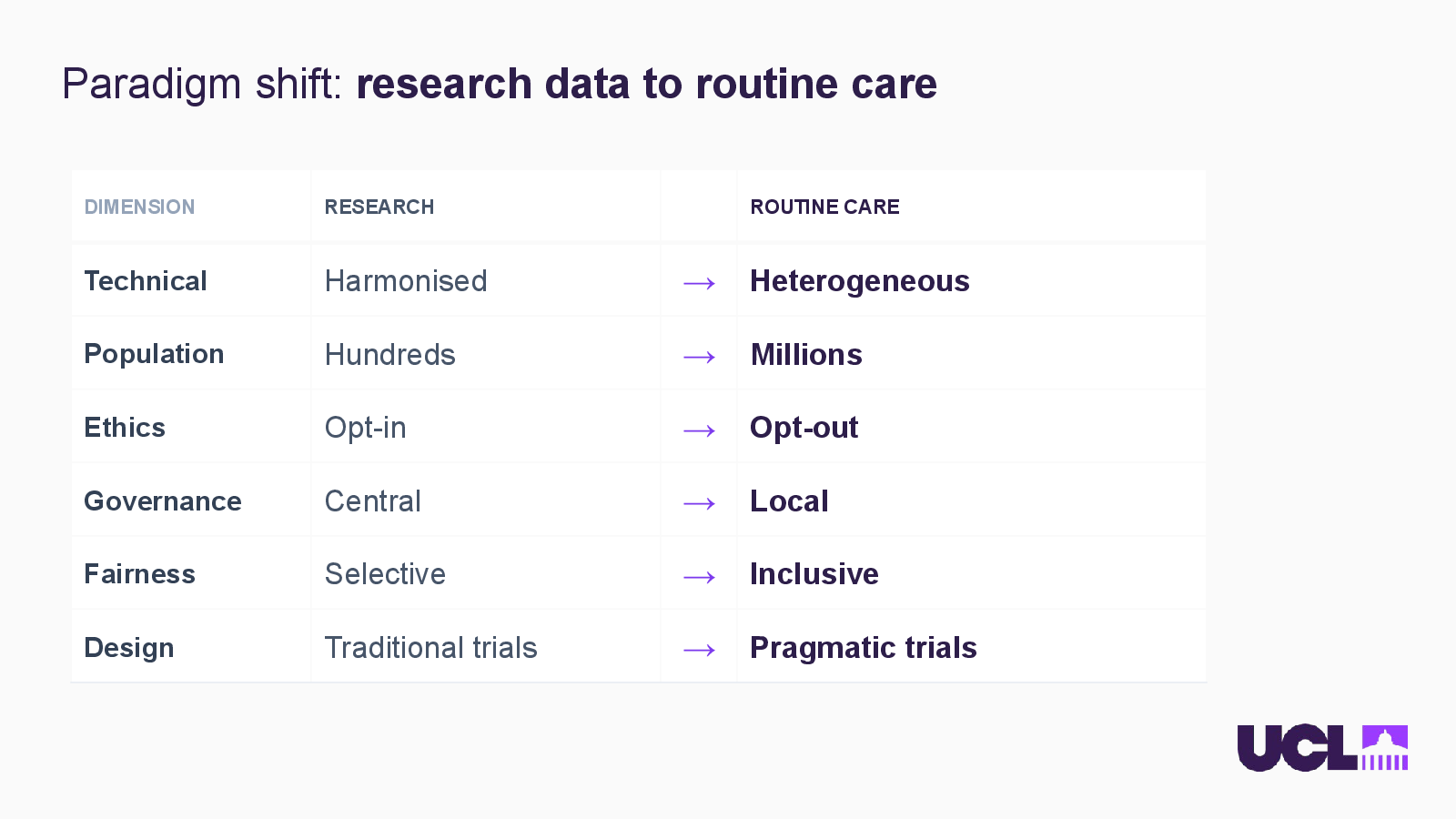
    Paradigm shift: research data to routine care
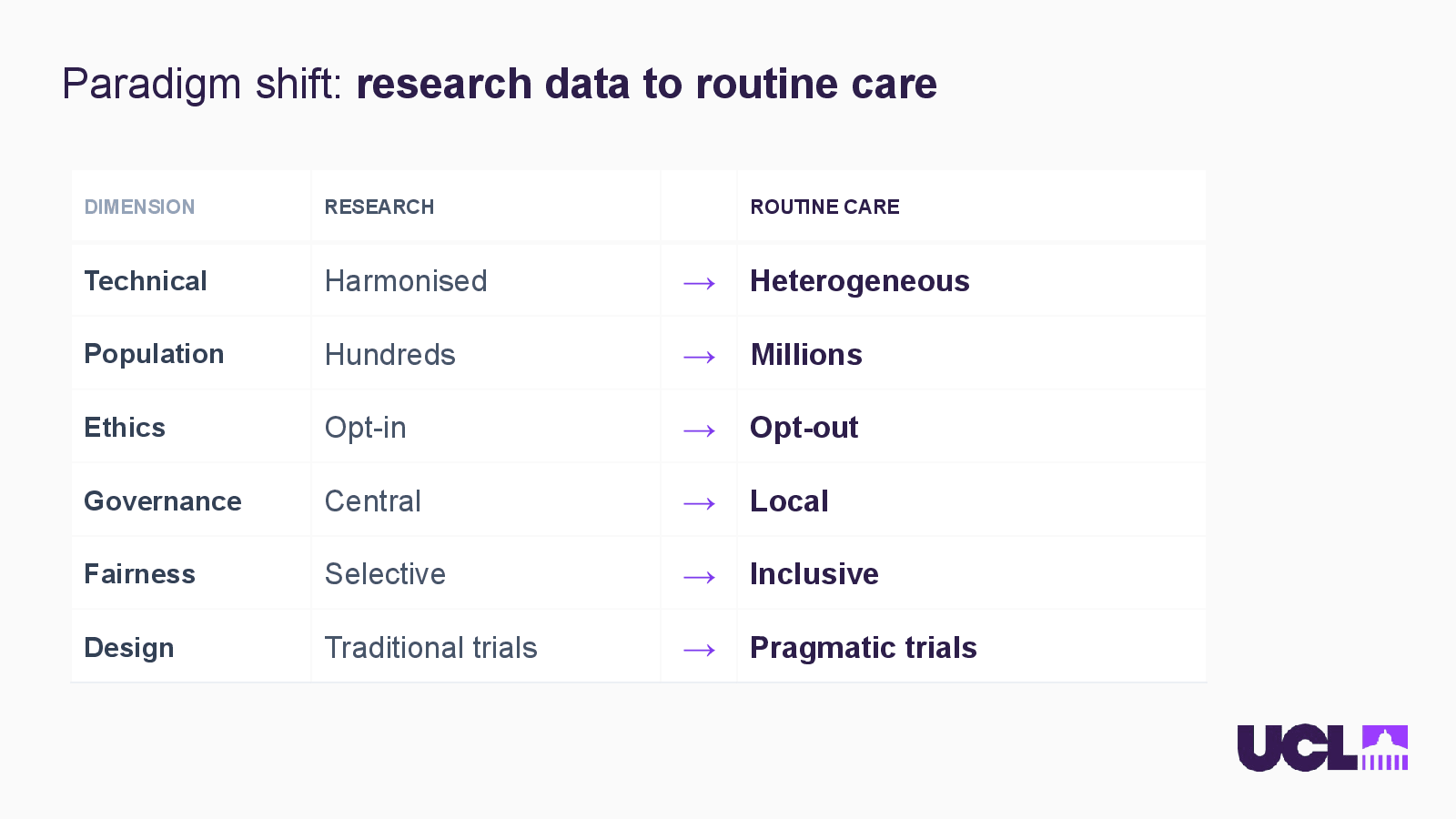
    Slide 6: Paradigm shift: research data to routine careOpen full-size slide
    • DIMENSION — RESEARCH — ROUTINE CARE
    • Technical — Harmonised — → — Heterogeneous
    • Population — Hundreds — → — Millions
    • Ethics — Opt-in — → — Opt-out
    • Governance — Central — → — Local
    • Fairness — Selective — → — Inclusive
    • Design — Traditional trials — → — Pragmatic trials
  7. Slide 07

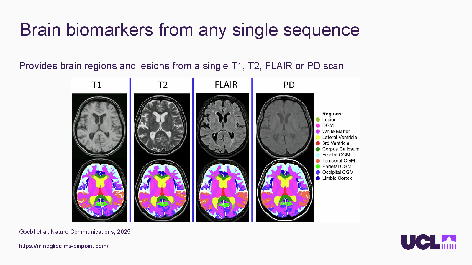
    Brain biomarkers from any single sequence
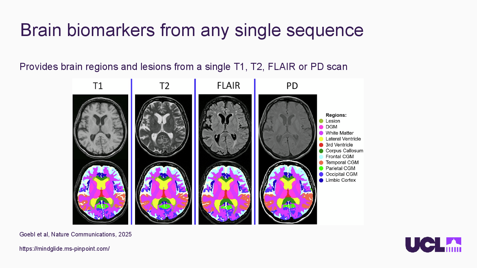
    Slide 7: Brain biomarkers from any single sequenceOpen full-size slide

    Provides brain regions and lesions from a single T1, T2, FLAIR or PD scan

    Goebl et al, Nature Communications, 2025

    https://mindglide.ms-pinpoint.com/

  8. Slide 08

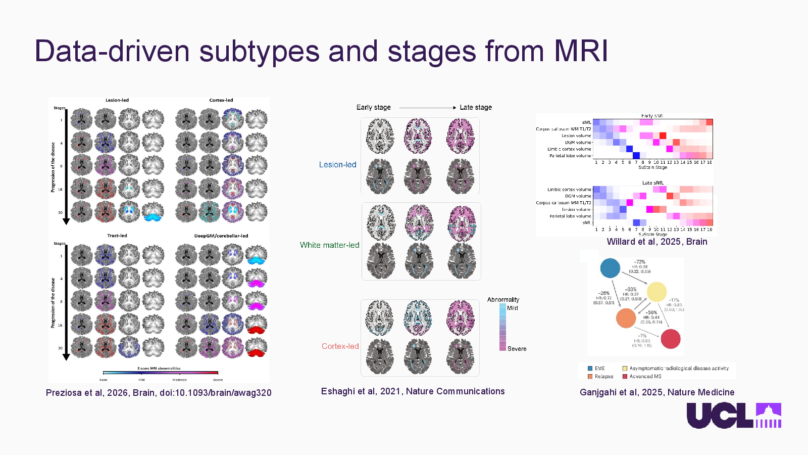
    Data-driven subtypes and stages from MRI
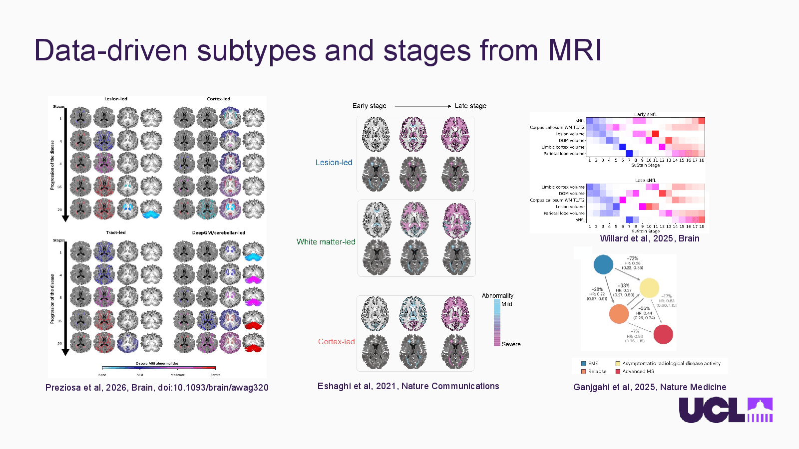
    Slide 8: Data-driven subtypes and stages from MRIOpen full-size slide

    Willard et al, 2025, Brain

    Preziosa et al, 2026, Brain, doi:10.1093/brain/awag320

    Eshaghi et al, 2021, Nature Communications

    Ganjgahi et al, 2025, Nature Medicine

    References

  9. Slide 09

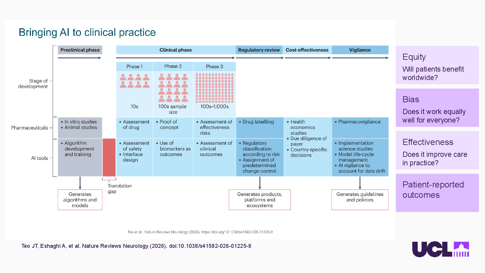
    Bringing AI to clinical practice
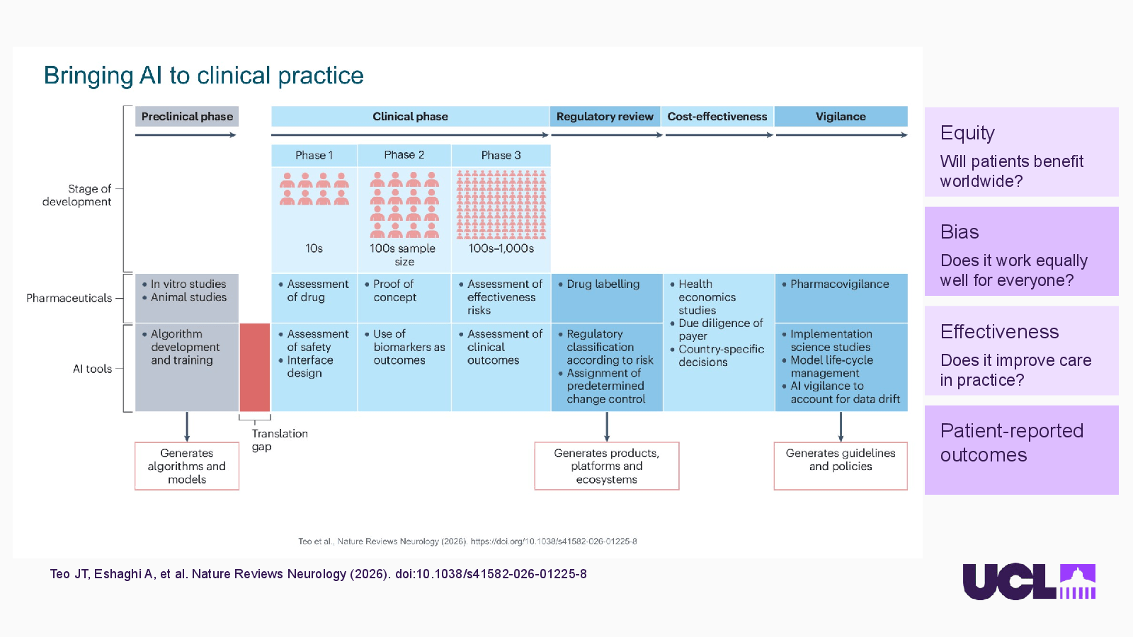
    Slide 9: Bringing AI to clinical practiceOpen full-size slide

    Teo JT, Eshaghi A, et al. Nature Reviews Neurology (2026). doi:10.1038/s41582-026-01225-8

    Equity

    Will patients benefit worldwide?

    Bias

    Does it work equally well for everyone?

    Effectiveness

    Does it improve care in practice?

    Patient-reported outcomes

    References

  10. Slide 10

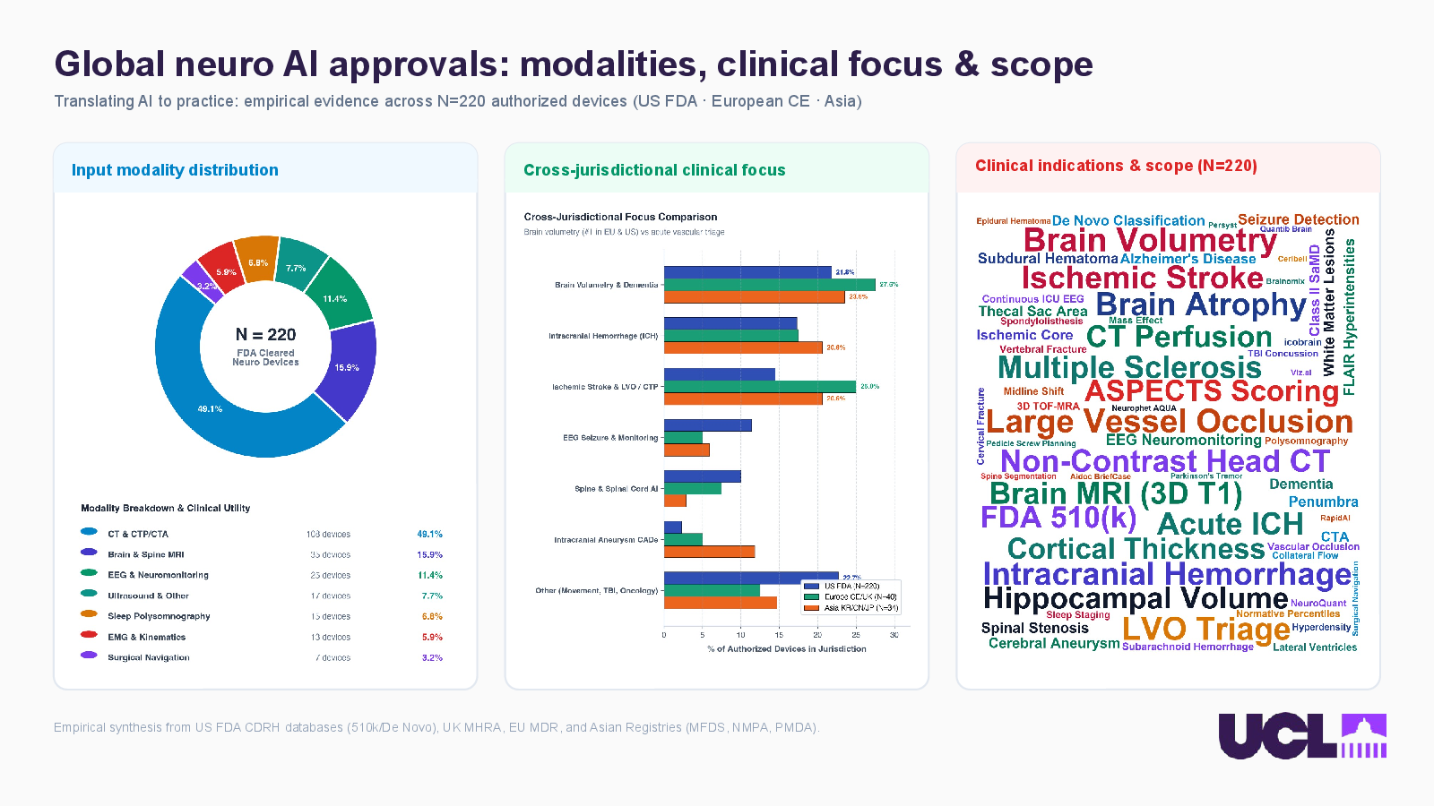
    Global neuro AI approvals: modalities, clinical focus & scope
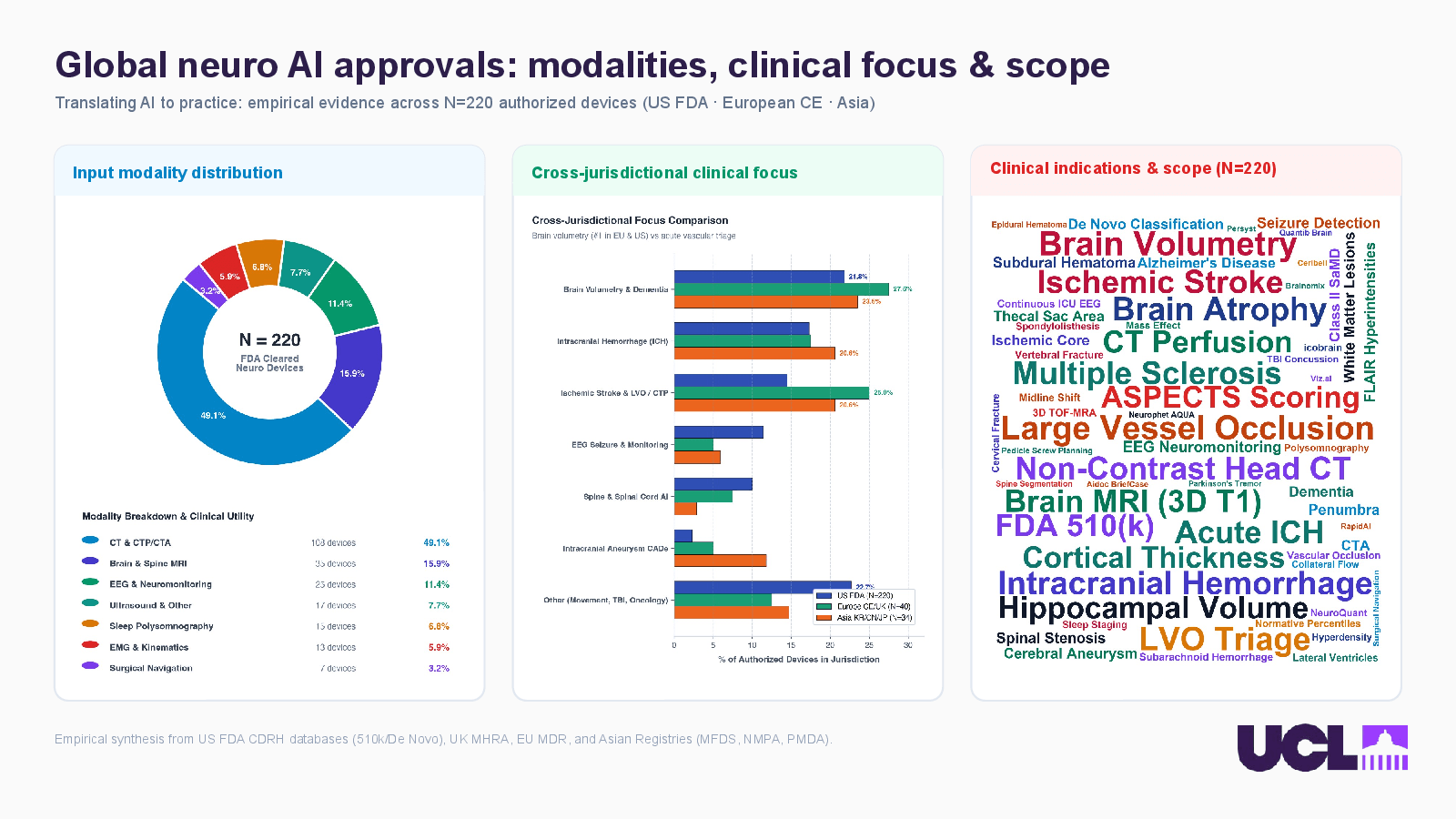
    Slide 10: Global neuro AI approvals: modalities, clinical focus & scopeOpen full-size slide

    Translating AI to practice: empirical evidence across N=220 authorized devices (US FDA · European CE · Asia)

    Input modality distribution

    Cross-jurisdictional clinical focus

    Clinical indications & scope (N=220)

    Empirical synthesis from US FDA CDRH databases (510k/De Novo), UK MHRA, EU MDR, and Asian Registries (MFDS, NMPA, PMDA).

  11. Slide 11

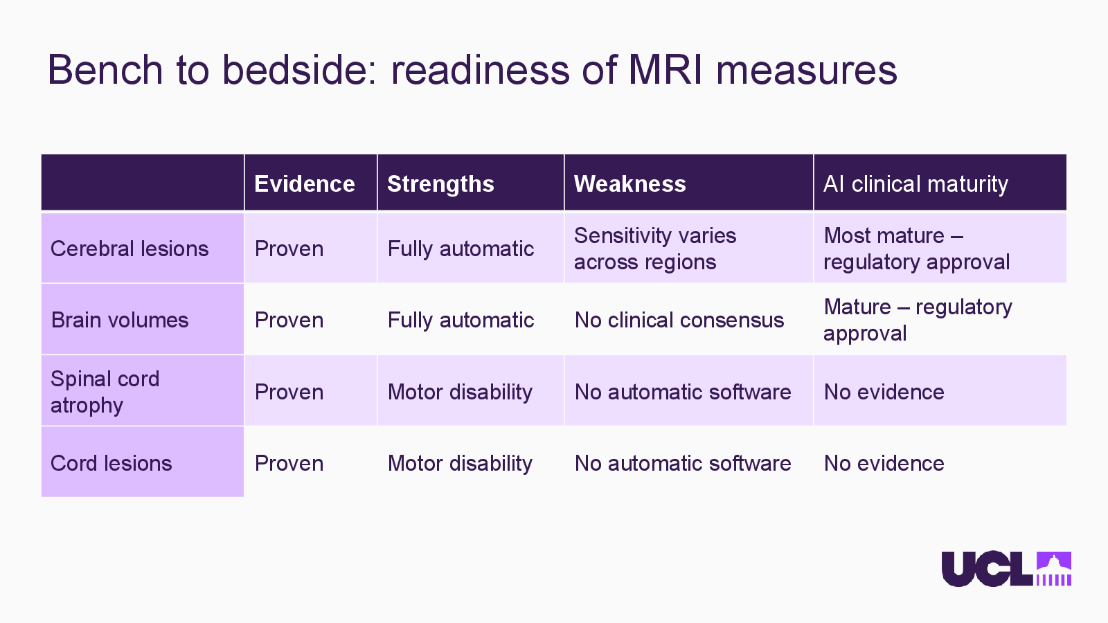
    Bench to bedside: readiness of MRI measures
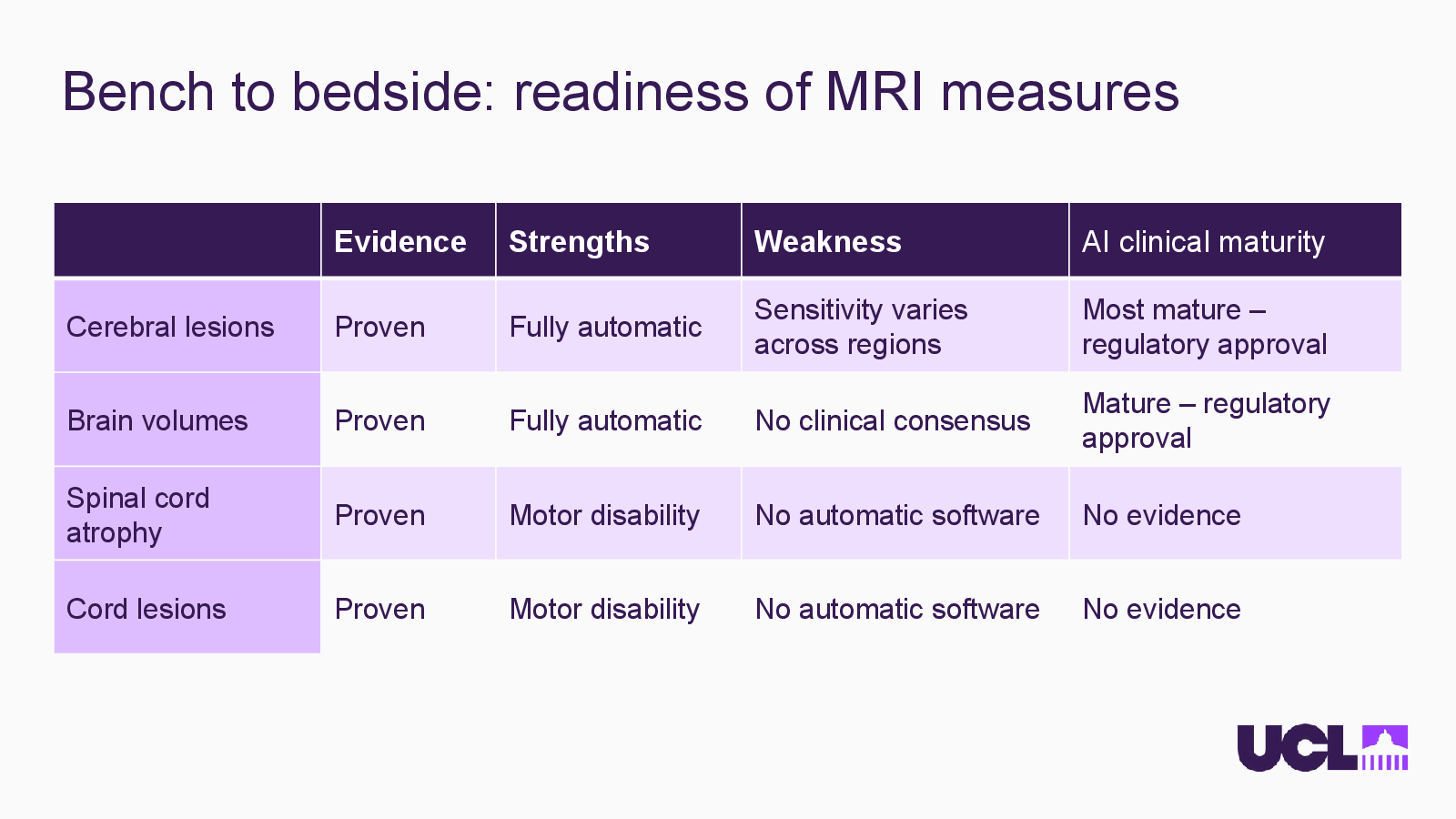
    Slide 11: Bench to bedside: readiness of MRI measuresOpen full-size slide
    • Evidence — Strengths — Weakness — AI clinical maturity
    • Cerebral lesions — Proven — Fully automatic — Sensitivity varies across regions — Most mature – regulatory approval
    • Brain volumes — Proven — Fully automatic — No clinical consensus — Mature – regulatory approval
    • Spinal cord atrophy — Proven — Motor disability — No automatic software — No evidence
    • Cord lesions — Proven — Motor disability — No automatic software — No evidence
  12. Slide 13

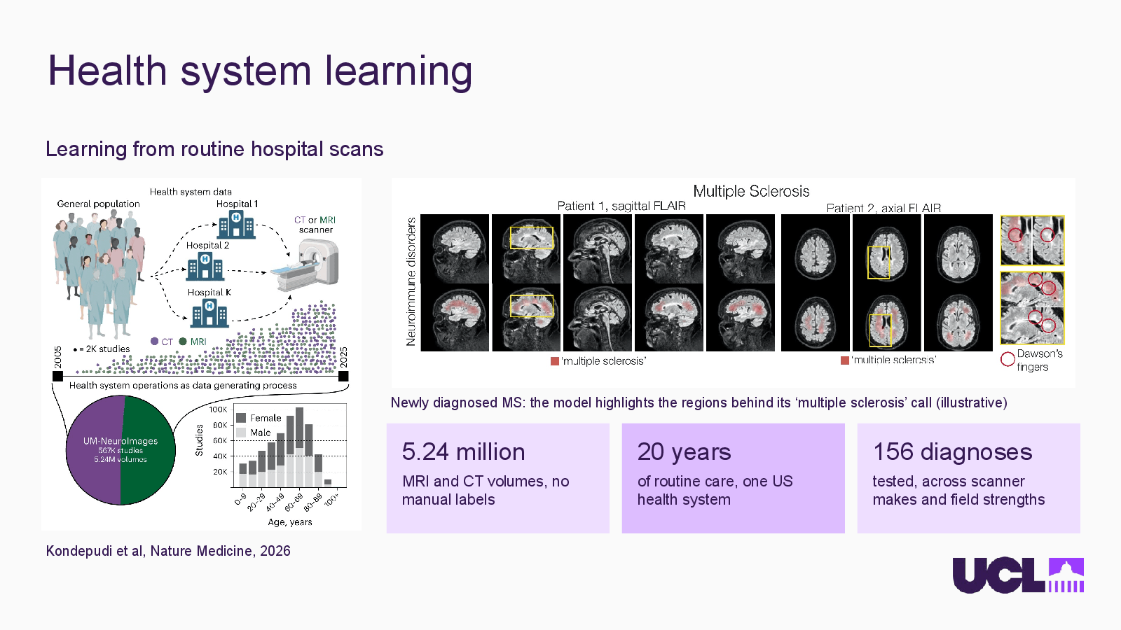
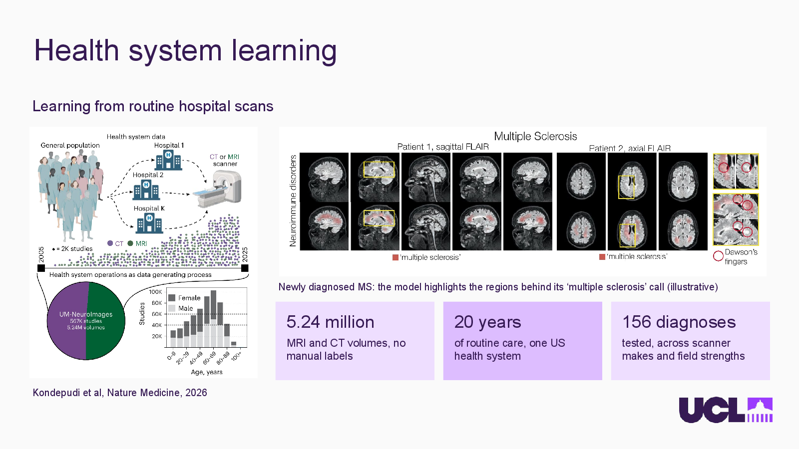
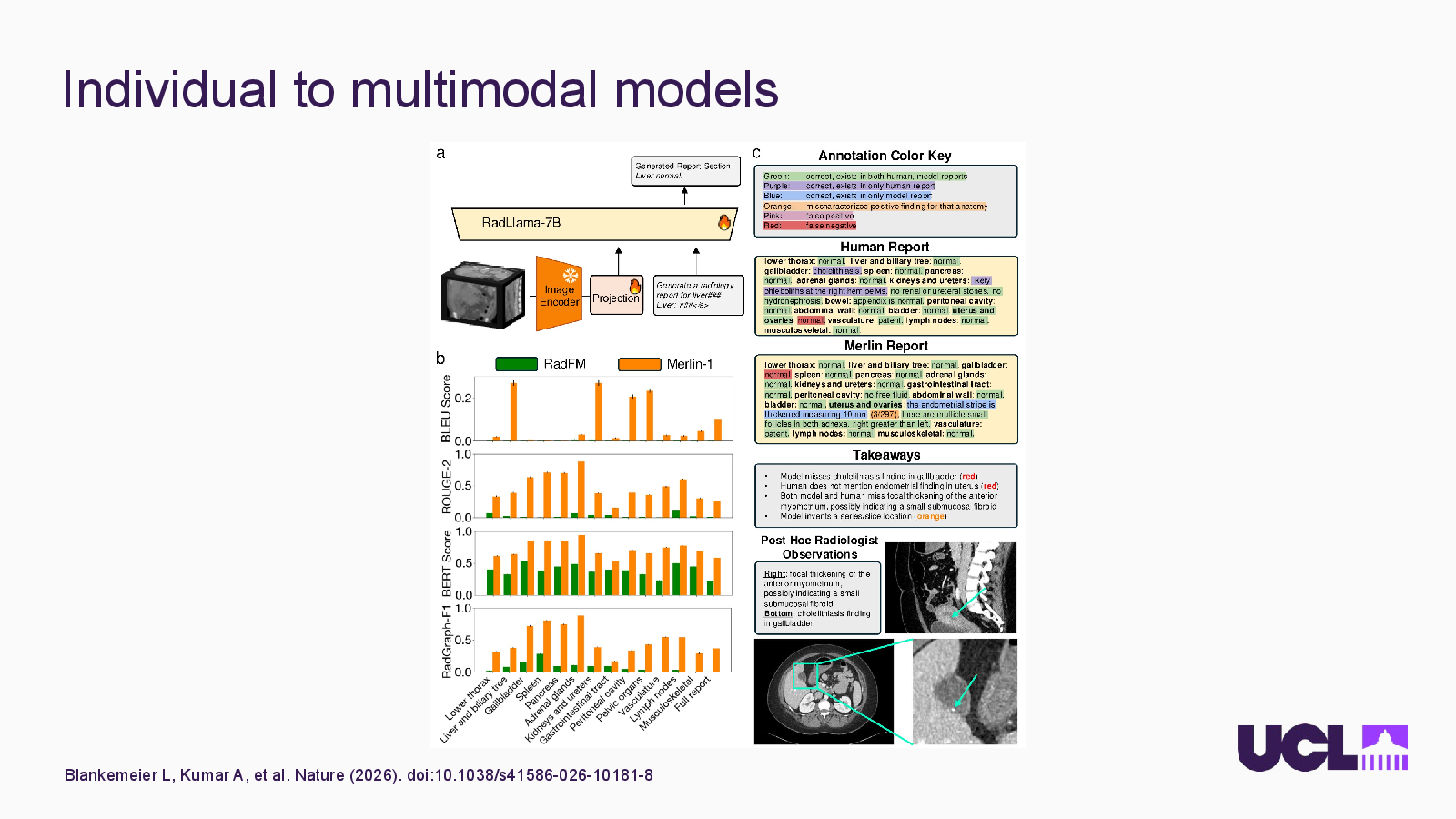
    Health system learning

    Slide 13: Health system learningOpen full-size slide

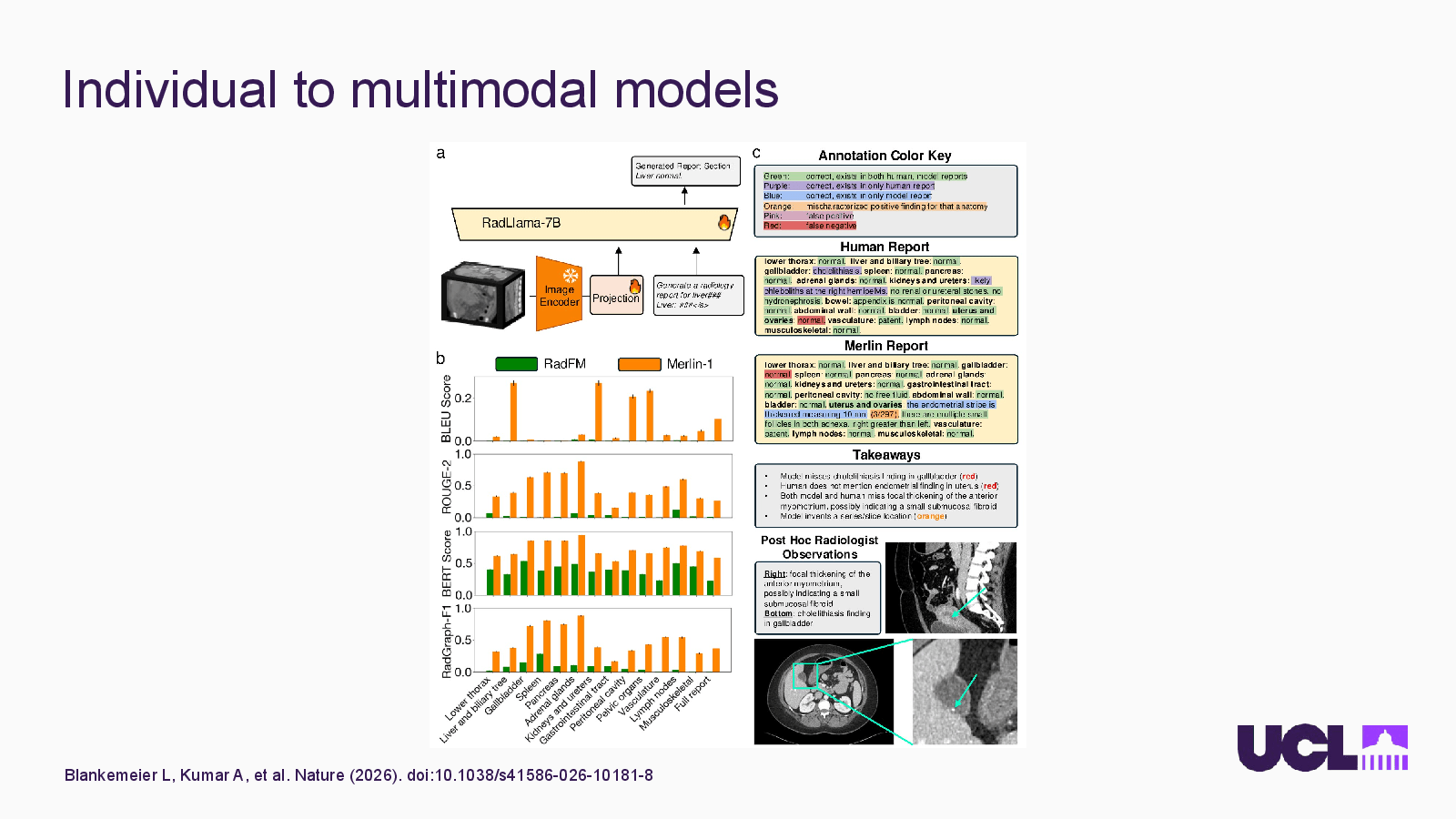
    Learning from routine hospital scans

    Newly diagnosed MS: the model highlights the regions behind its ‘multiple sclerosis’ call (illustrative)

    5.24 million

    MRI and CT volumes, no manual labels

    20 years

    of routine care, one US health system

    156 diagnoses

    tested, across scanner makes and field strengths

    Kondepudi et al, Nature Medicine, 2026

  13. Slide 14

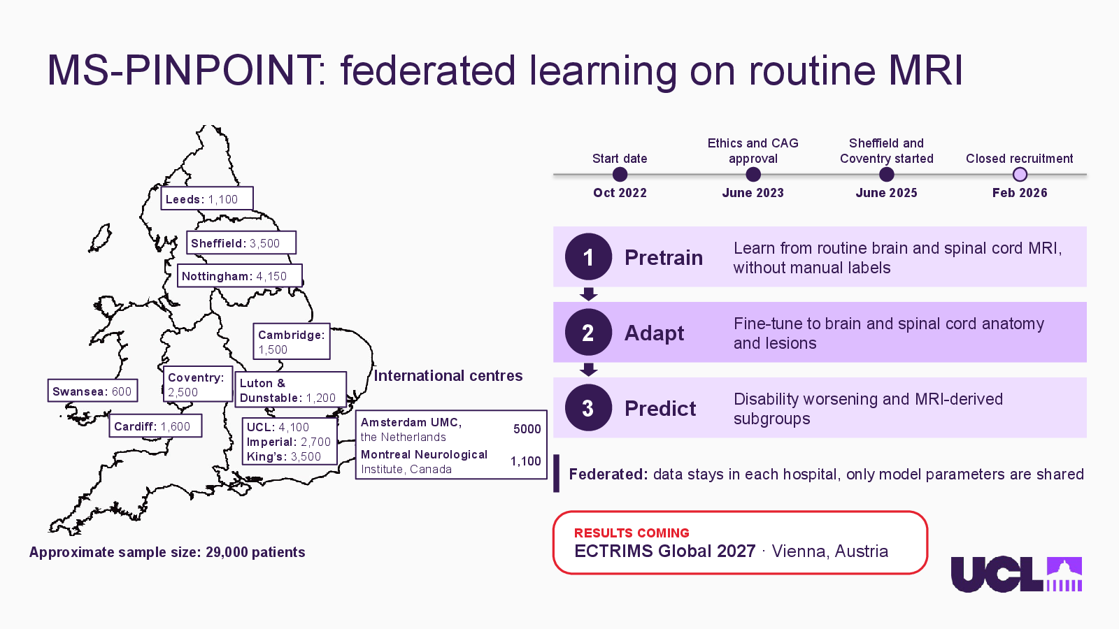
    MS-PINPOINT: federated learning on routine MRI
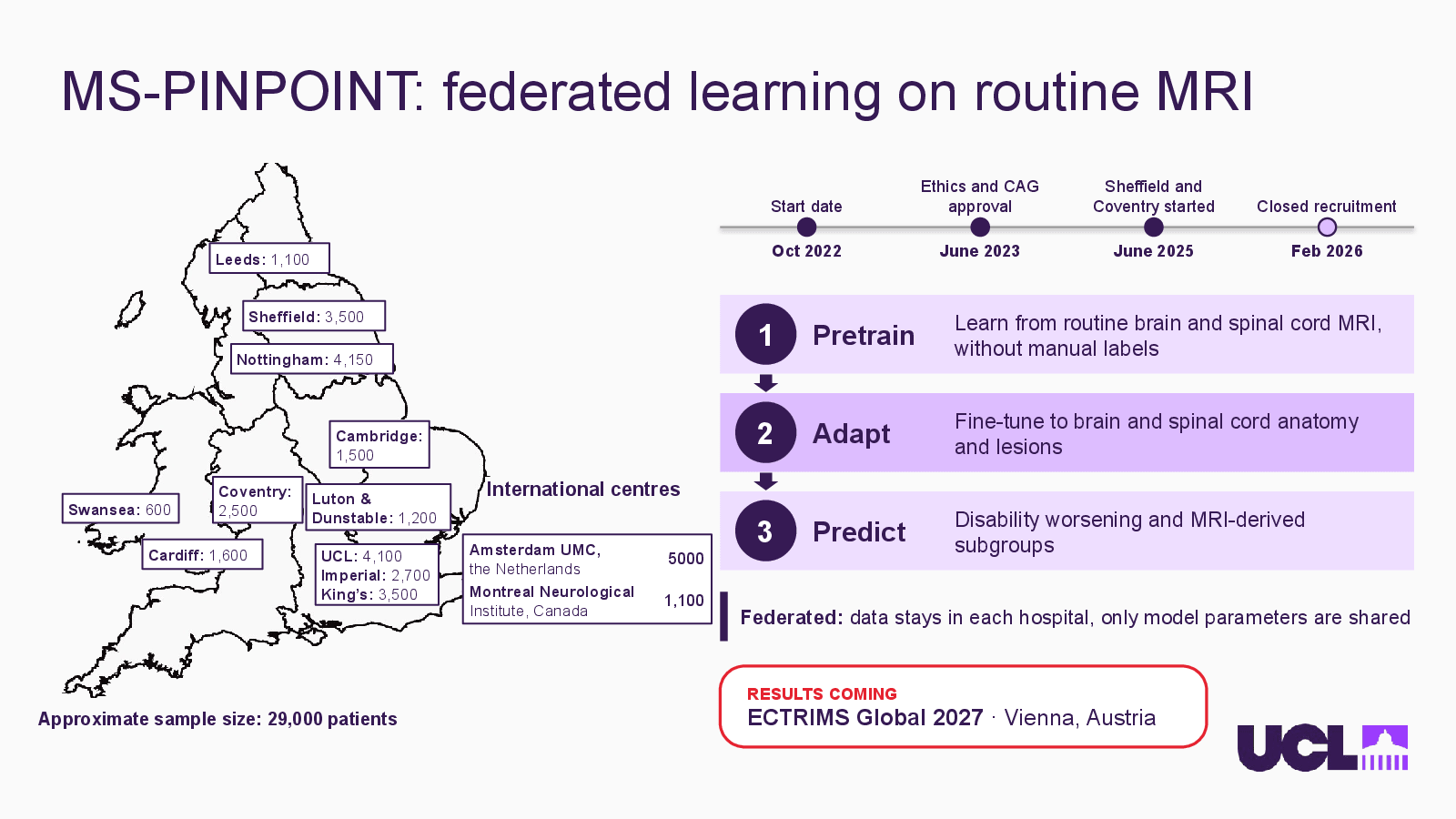
    Slide 14: MS-PINPOINT: federated learning on routine MRIOpen full-size slide

    Start date

    Ethics and CAG approval

    Sheffield and Coventry started

    Closed recruitment

    Oct 2022

    June 2023

    June 2025

    Feb 2026

    Leeds: 1,100

    Pretrain

    Learn from routine brain and spinal cord MRI, without manual labels

    Sheffield: 3,500

    Nottingham: 4,150

    Adapt

    Fine-tune to brain and spinal cord anatomy and lesions

    Cambridge:

    1,500

    International centres

    Coventry:

    2,500

    Luton &

    Dunstable: 1,200

    Predict

    Disability worsening and MRI-derived subgroups

    Swansea: 600

    Amsterdam UMC,

    the Netherlands

    5000

    Cardiff: 1,600

    UCL: 4,100

    Imperial: 2,700

    King’s: 3,500

    Montreal Neurological

    Institute, Canada

    1,100

    Federated: data stays in each hospital, only model parameters are shared

    RESULTS COMING

    ECTRIMS Global 2027 · Vienna, Austria

    Approximate sample size: 29,000 patients

  14. Slide 16

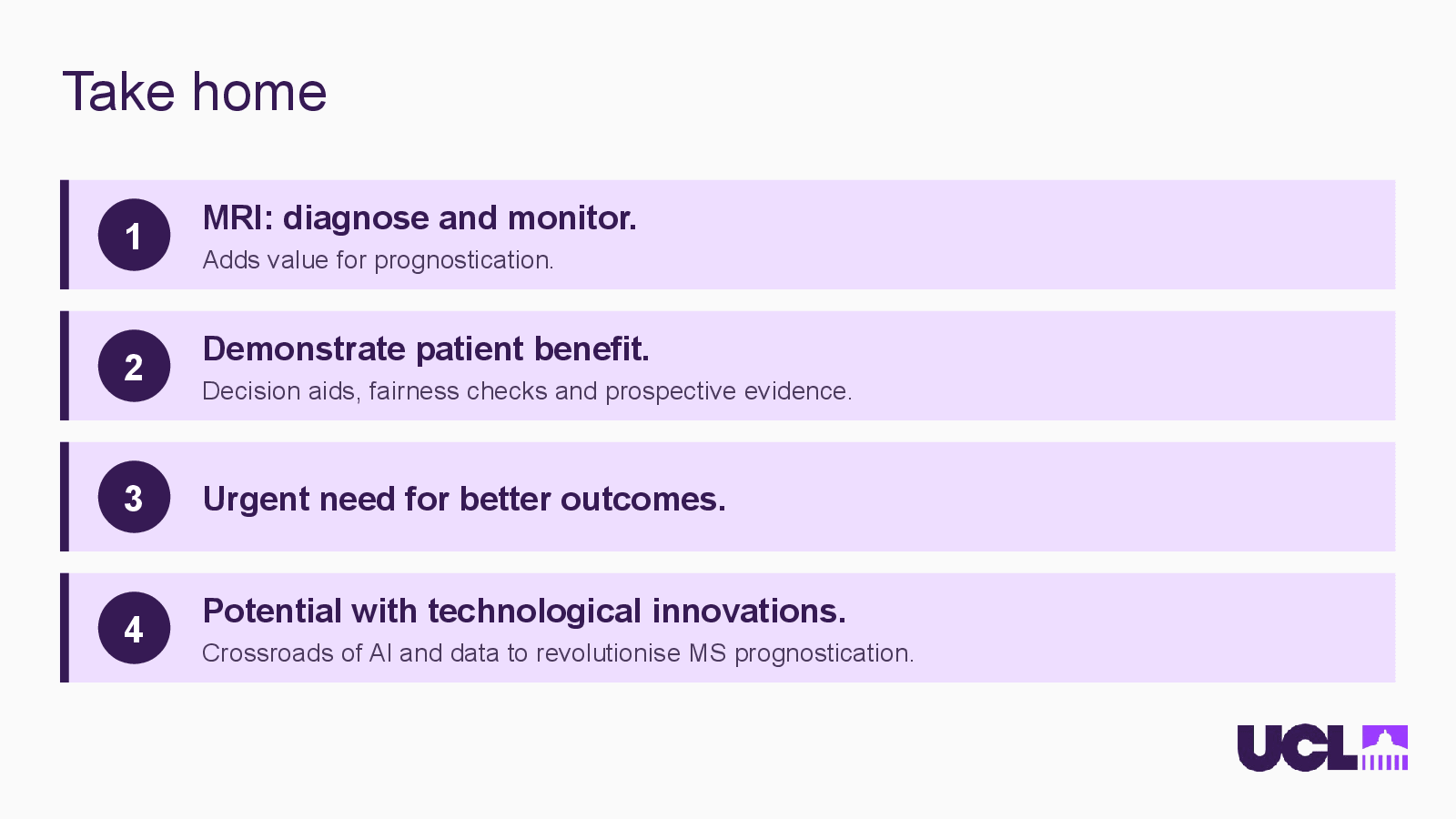
    Take home

    Slide 16: Take homeOpen full-size slide

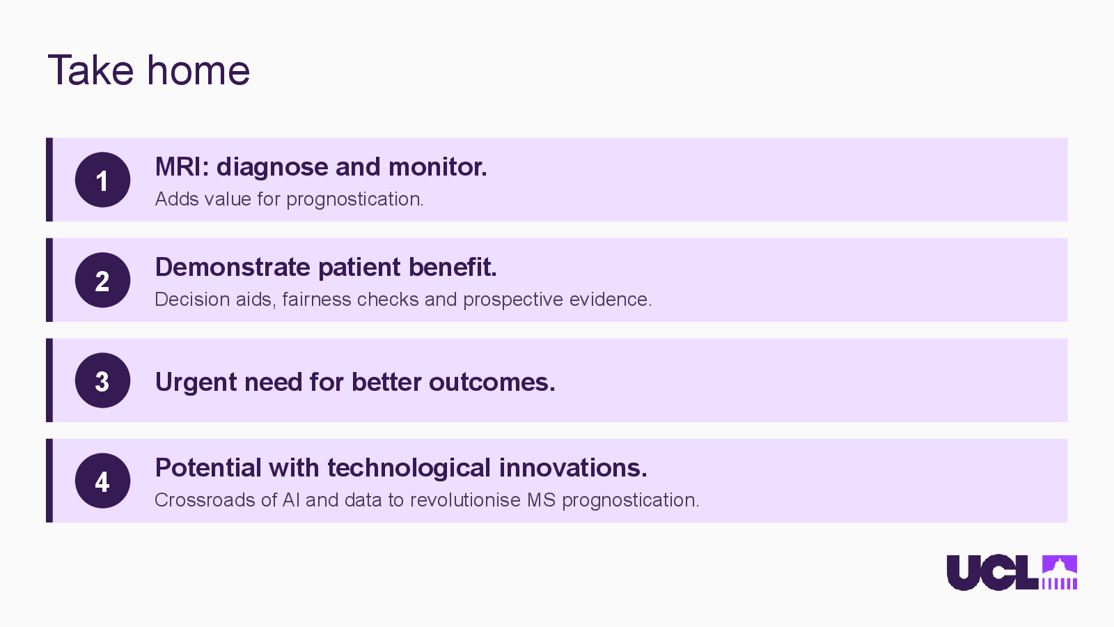
    MRI: diagnose and monitor.

    Adds value for prognostication.

    Demonstrate patient benefit.

    Decision aids, fairness checks and prospective evidence.

    Urgent need for better outcomes.

    Potential with technological innovations.

    Crossroads of AI and data to revolutionise MS prognostication.

  15. Slide 17

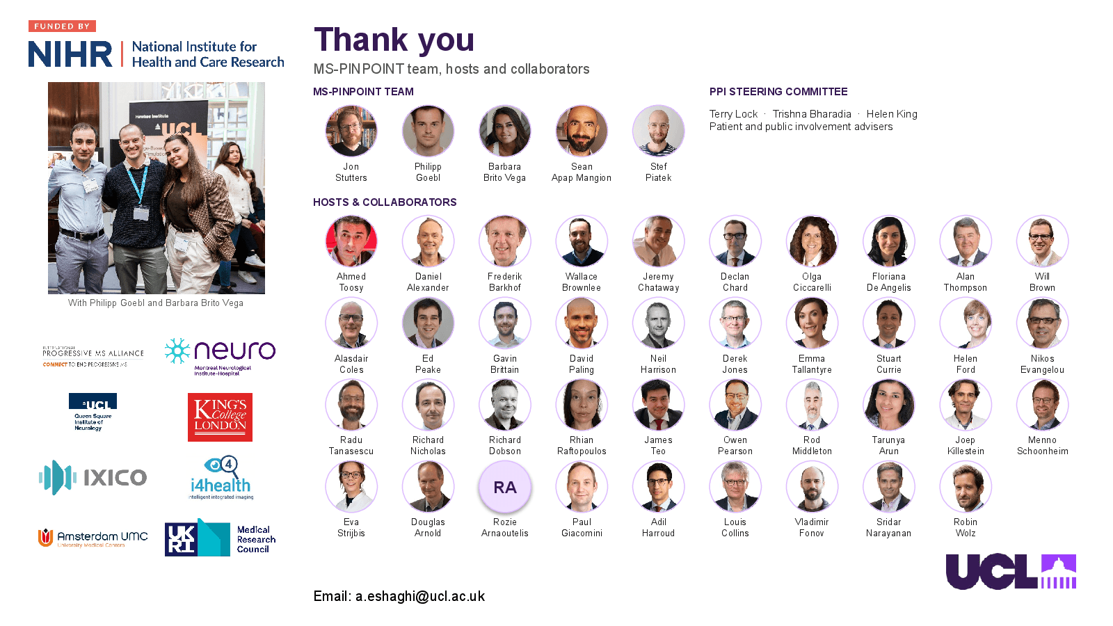
    Thank you

    Slide 17: Thank youOpen full-size slide

    Terry Lock · Trishna Bharadia · Helen King

    Thank you

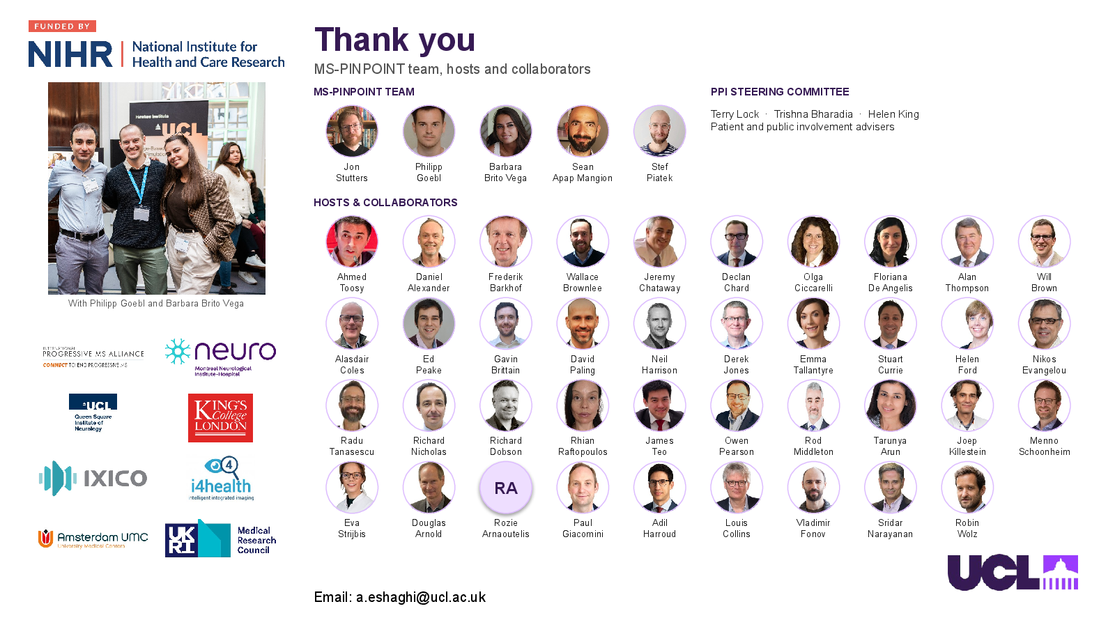
    MS-PINPOINT team, hosts and collaborators

    MS-PINPOINT TEAM

    PPI STEERING COMMITTEE

    Patient and public involvement advisers

    Jon

    Stutters

    Philipp

    Goebl

    Barbara

    Brito Vega

    Sean

    Apap Mangion

    Stef

    Piatek

    HOSTS & COLLABORATORS

    Ahmed

    Toosy

    Daniel

    Alexander

    Frederik

    Barkhof

    Wallace

    Brownlee

    Jeremy

    Chataway

    Declan

    Chard

    Olga

    Ciccarelli

    Floriana

    De Angelis

    Alan

    Thompson

    Will

    Brown

    With Philipp Goebl and Barbara Brito Vega

    Alasdair

    Coles

    Ed

    Peake

    Gavin

    Brittain

    David

    Paling

    Neil

    Harrison

    Derek

    Jones

    Emma

    Tallantyre

    Stuart

    Currie

    Helen

    Ford

    Nikos

    Evangelou

    Radu

    Tanasescu

    Richard

    Nicholas

    Dobson

    Rhian

    Raftopoulos

    James

    Teo

    Owen

    Pearson

    Rod

    Middleton

    Tarunya

    Arun

    Joep

    Killestein

    Menno

    Schoonheim

    RA

    Eva

    Strijbis

    Douglas

    Arnold

    Rozie

    Arnaoutelis

    Paul

    Giacomini

    Adil

    Harroud

    Louis

    Collins

    Vladimir

    Fonov

    Sridar

    Narayanan

    Robin

    Wolz

    Email: a.eshaghi@ucl.ac.uk