Workshop ·
Predicting multiple sclerosis progression with conventional MRI using artificial intelligence
What routine MRI can and cannot predict about MS progression, and where AI may add practical value.
41st MAGNIMS Plenary Meeting and Workshop · Van der Valk Hotel Amsterdam-Zuidas, Amsterdam
- Delivered
- Event
- 41st MAGNIMS Plenary Meeting and Workshop
- Place
- Van der Valk Hotel Amsterdam-Zuidas, Amsterdam
- Format
- Workshop
- Published deck
- 14 slides
From the slides
Deck notes
This workshop talk examined the use of conventional MRI to predict progression in multiple sclerosis. It balanced the value of routinely acquired images against their known limits as individual prognostic tools.
The question
Can artificial intelligence recover useful prognostic information from heterogeneous, routinely collected MRI without overstating what those scans can predict?
Deck outline
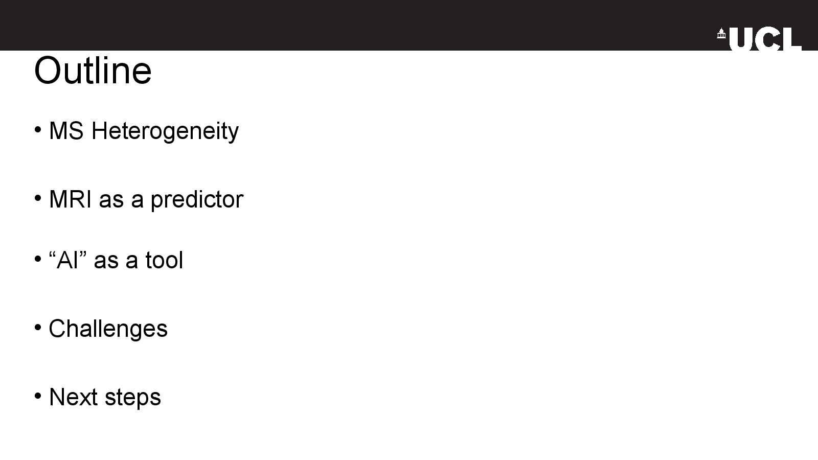
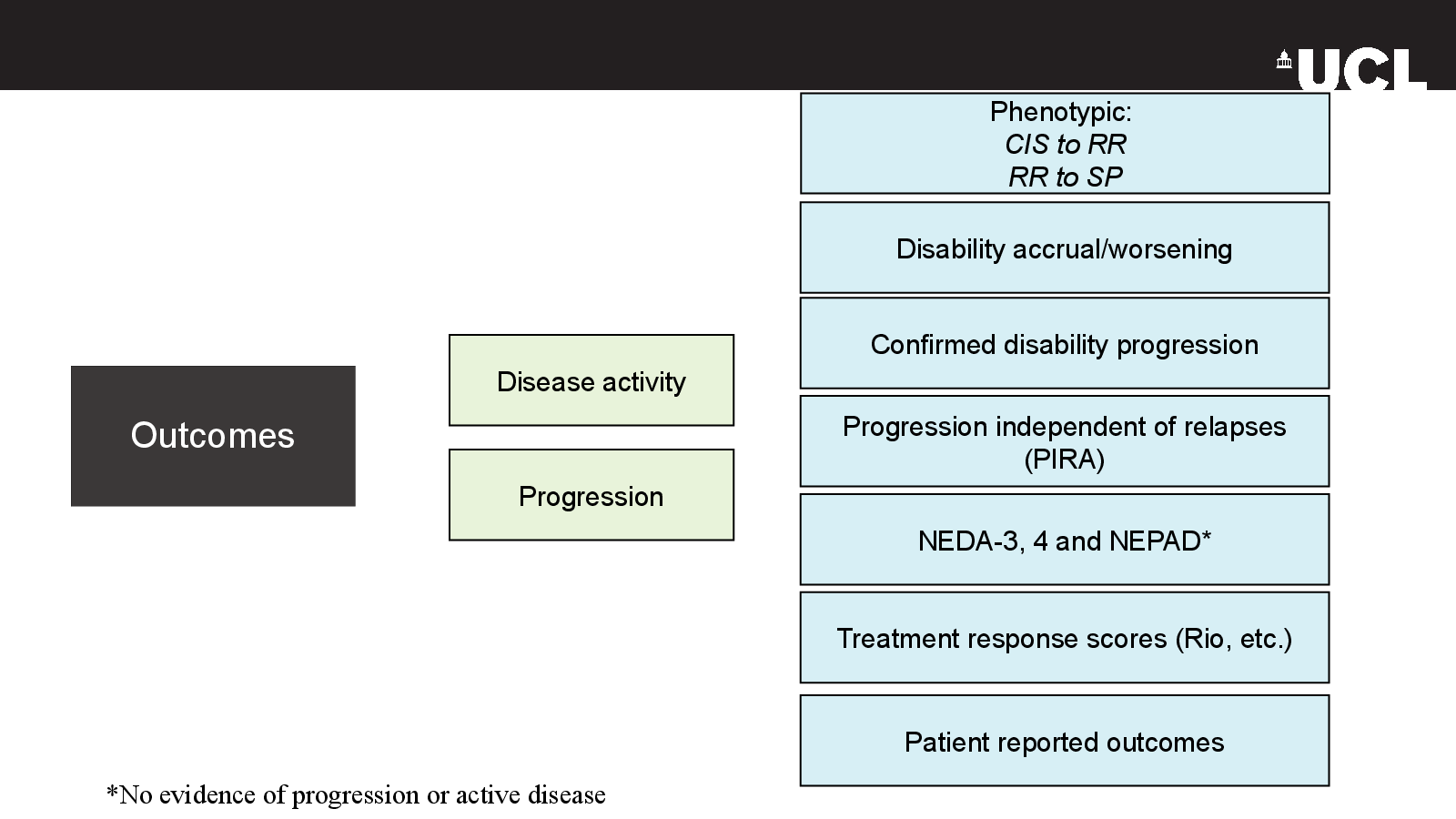
- Clinical definitions of disease activity, progression and disability worsening
- Conventional brain and spinal cord MRI markers
- AI analysis of heterogeneous clinical scans
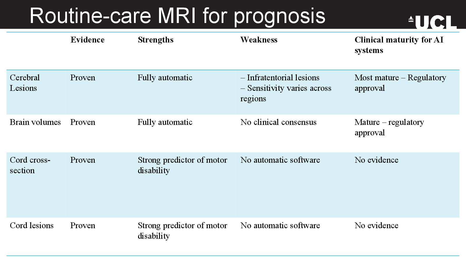
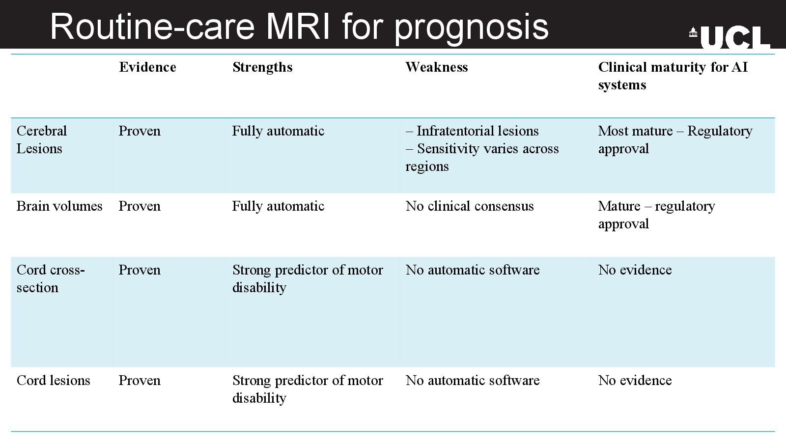
- Evidence, limitations and clinical maturity across imaging measures
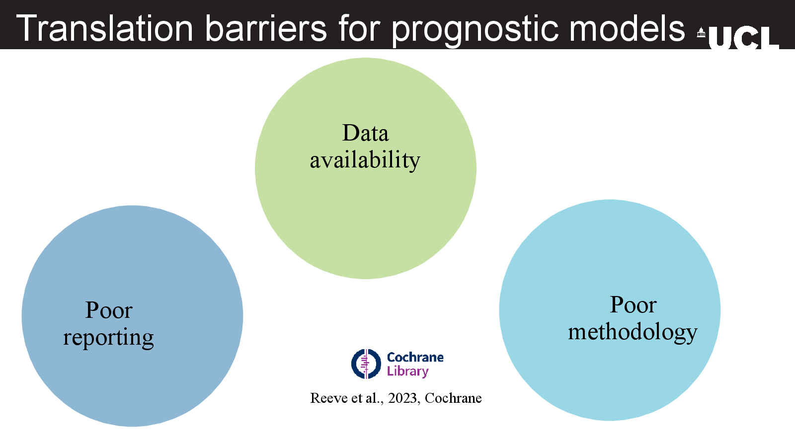
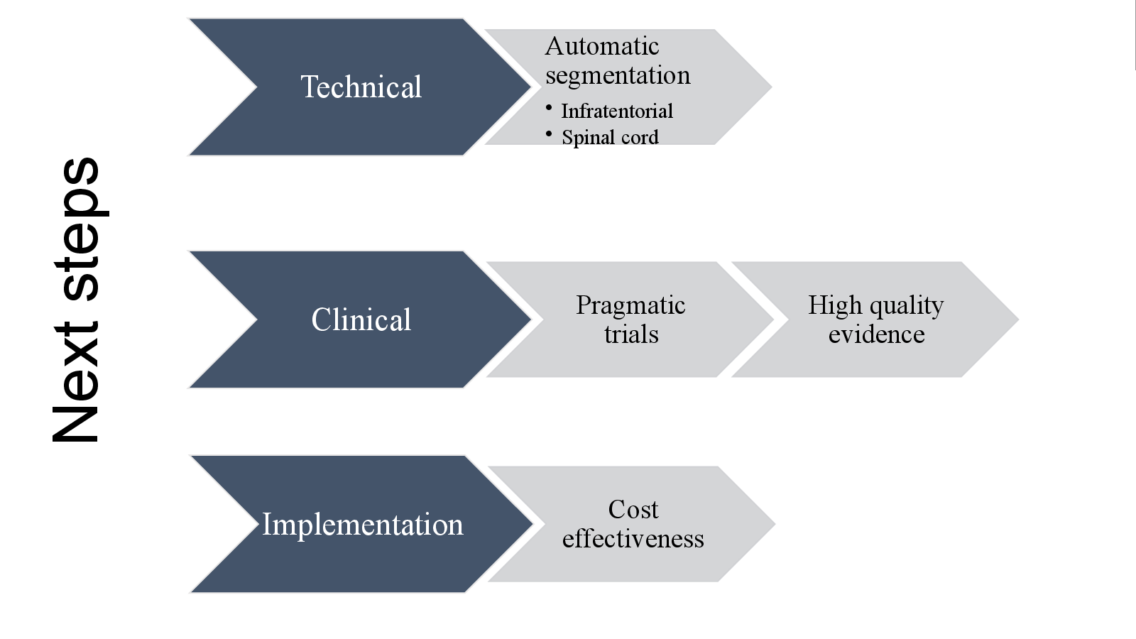
- Barriers to translating prognostic models into practice
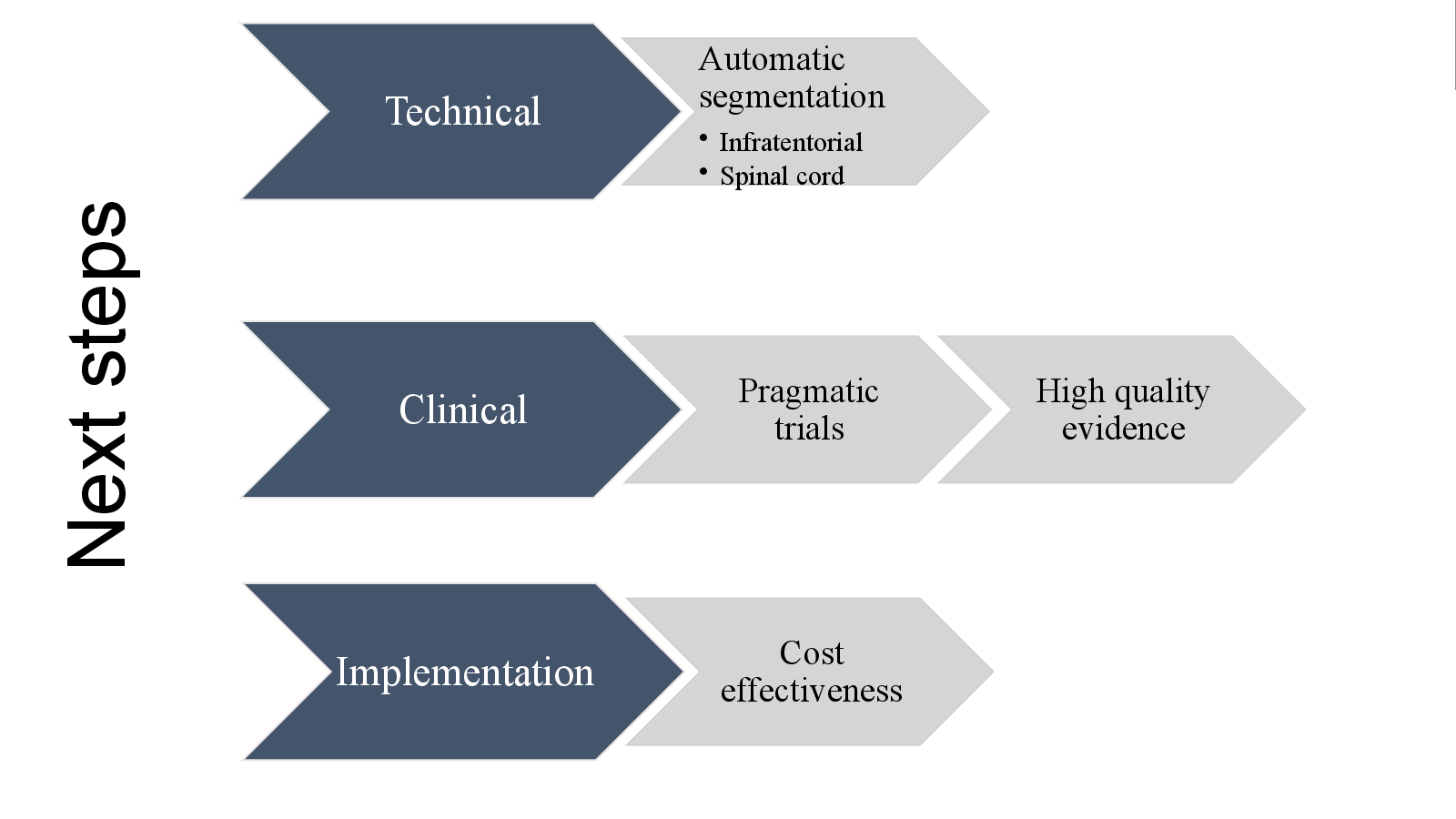
- Priorities for routine brain and spinal cord imaging
Take-away
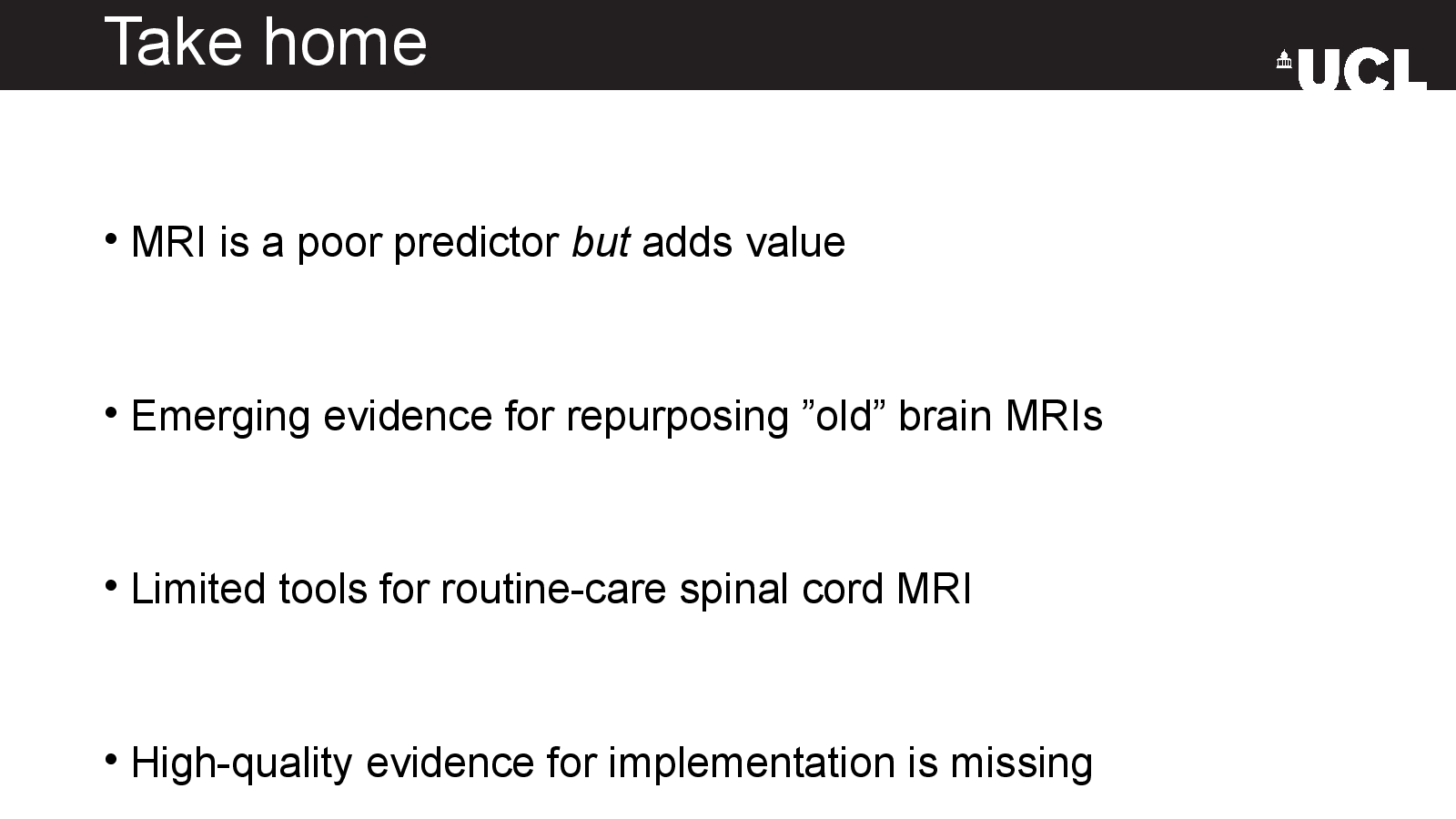
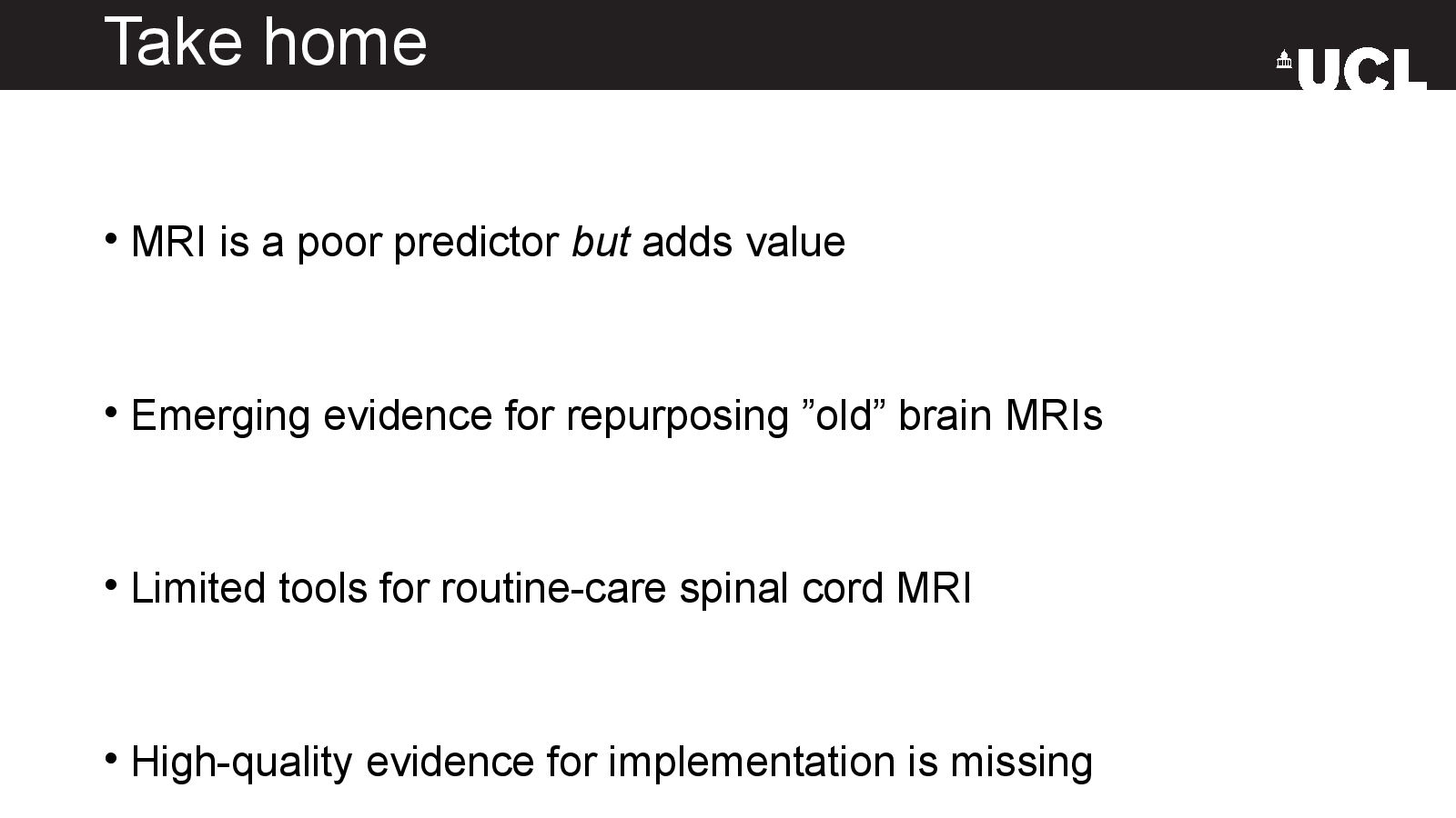
Conventional MRI is a limited predictor on its own, but it can add value when analysed carefully. Better spinal cord tools and high-quality implementation evidence remain important gaps.
Original deck
Slide by slide
Read the 14-slide presentation in order, or use the index to jump.
Slide 01
Predicting multiple sclerosis progression with conventional MRI using artificial intelligence
Slide 02
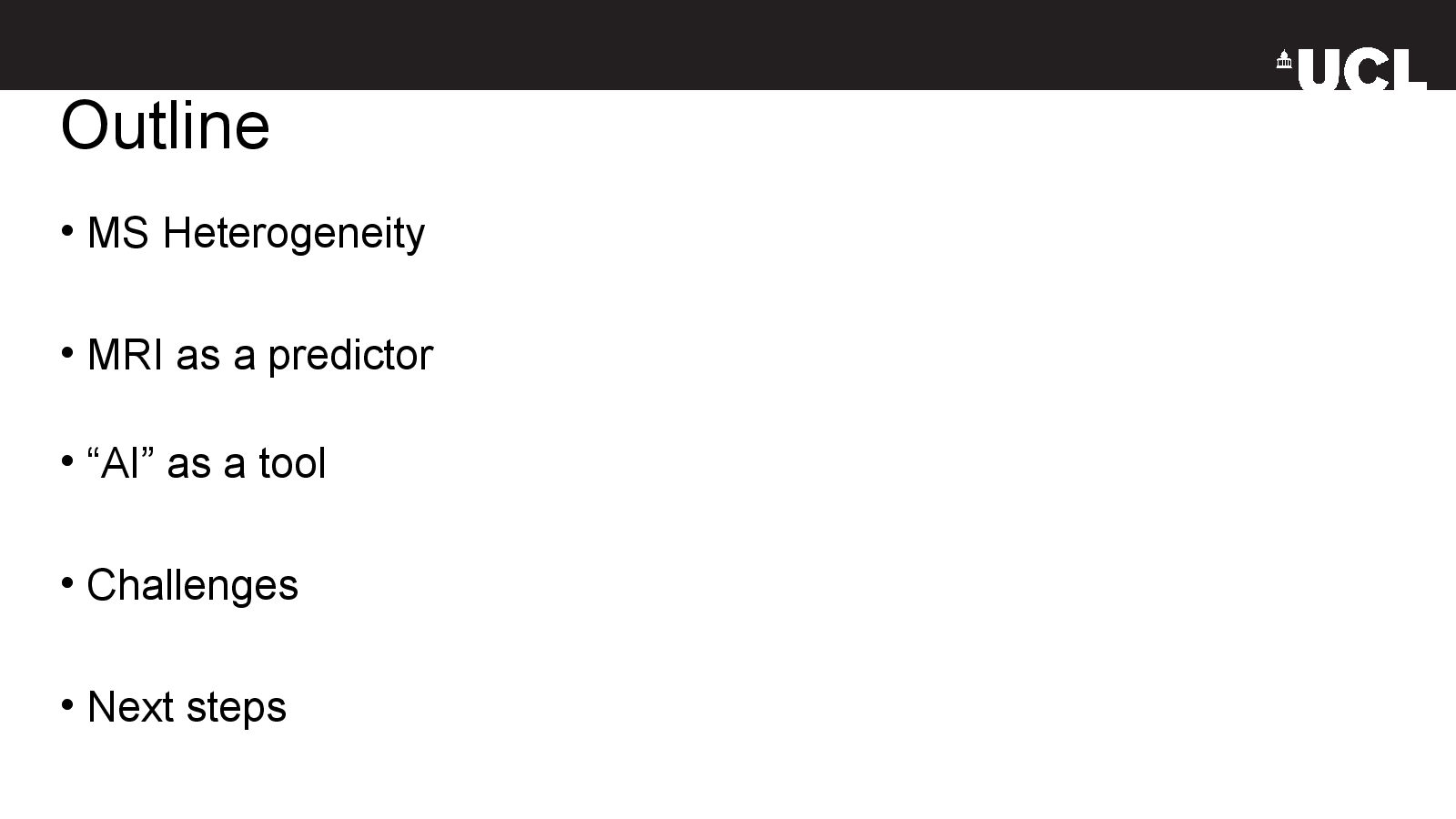
Outline
Slide 03
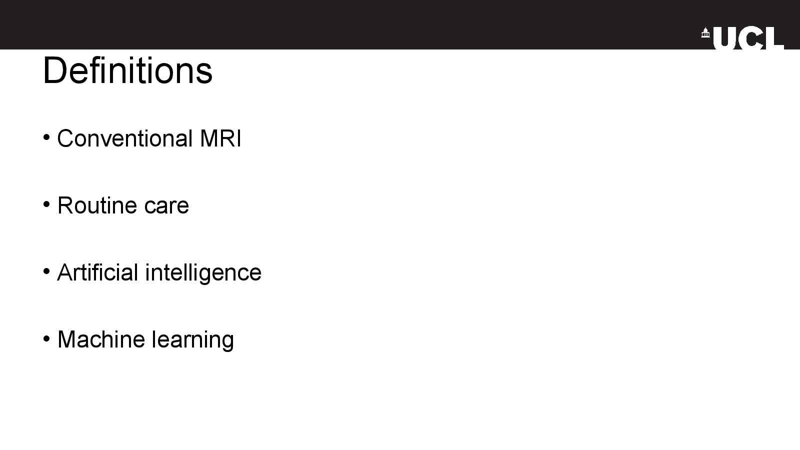
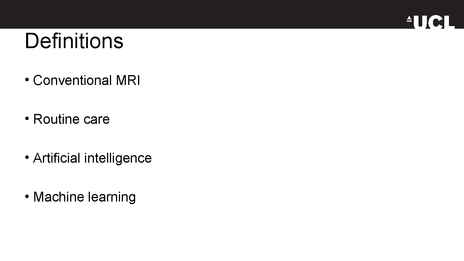
Definitions
Slide 04
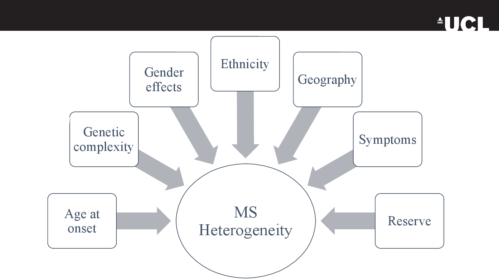
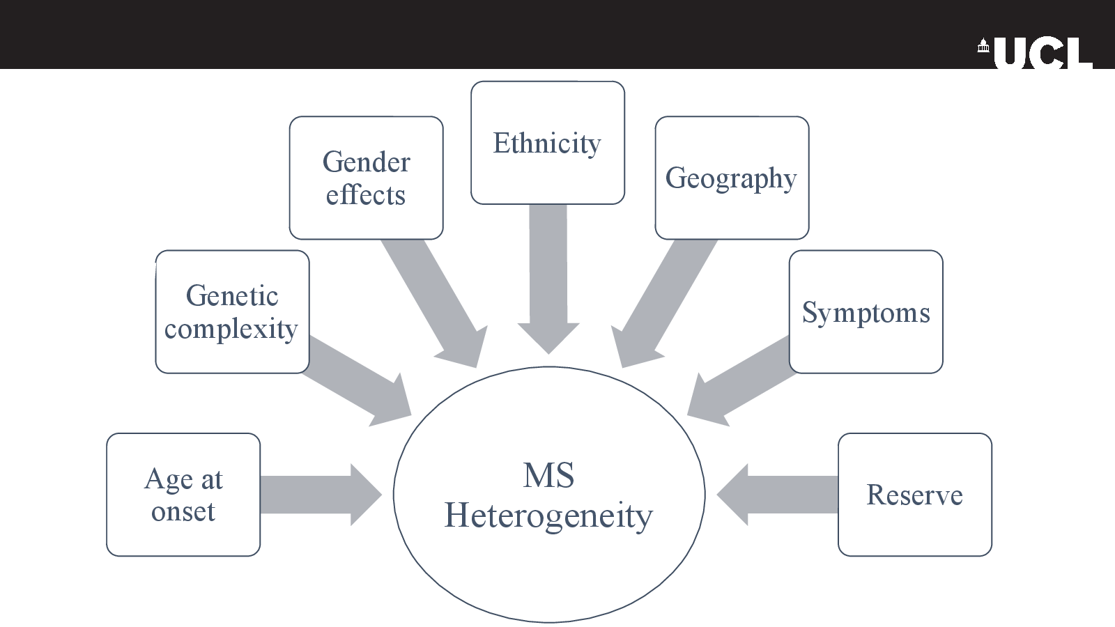
Slide 4
Slide 05
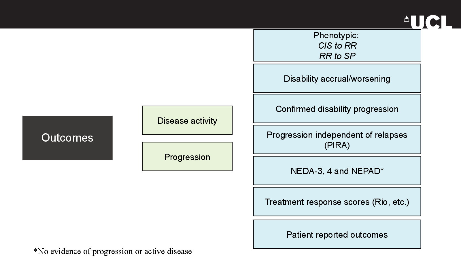
Outcomes
Slide 06
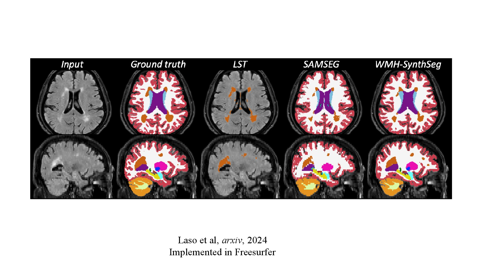
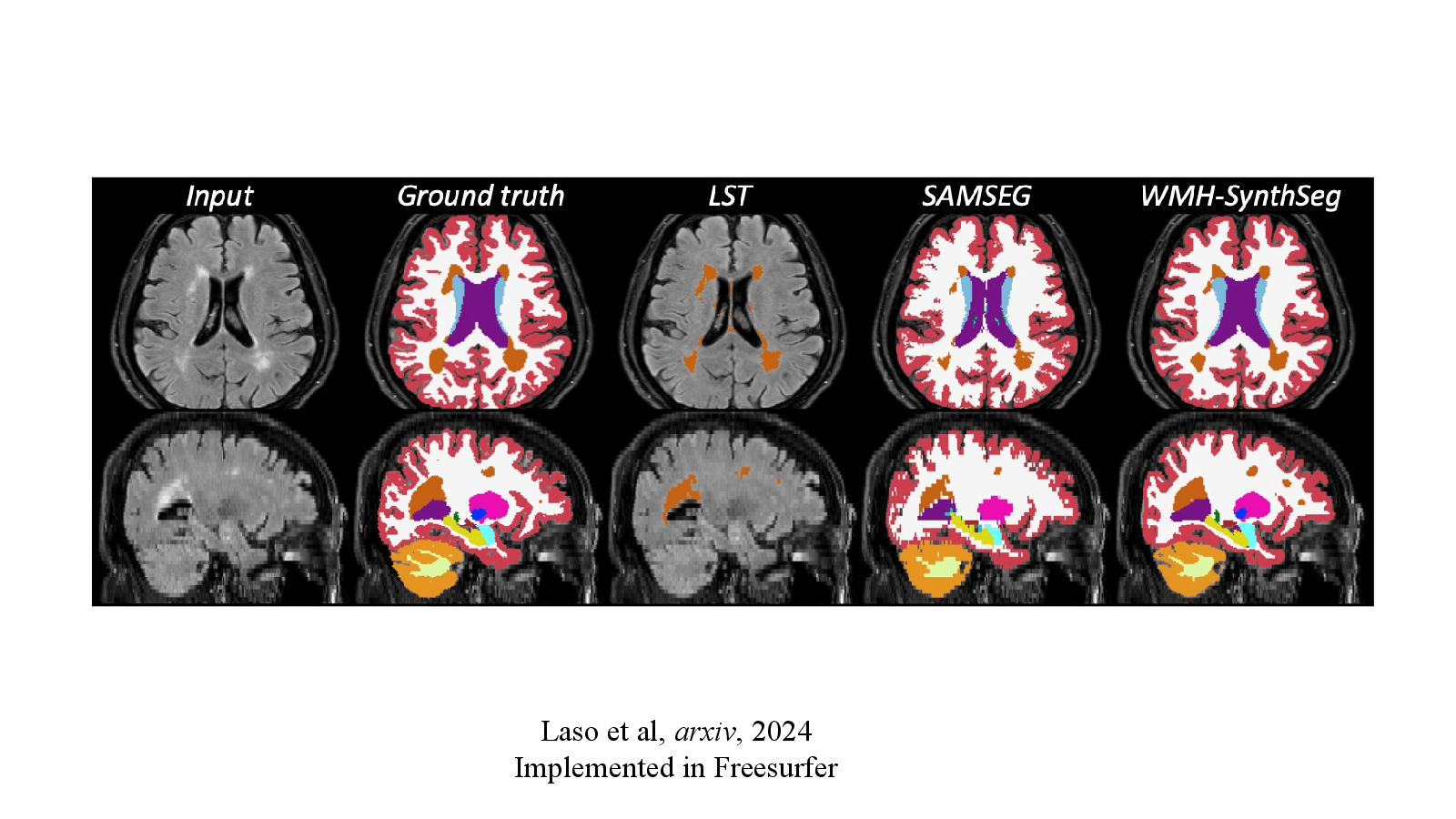
Laso et al, arxiv, 2024
Slide 07
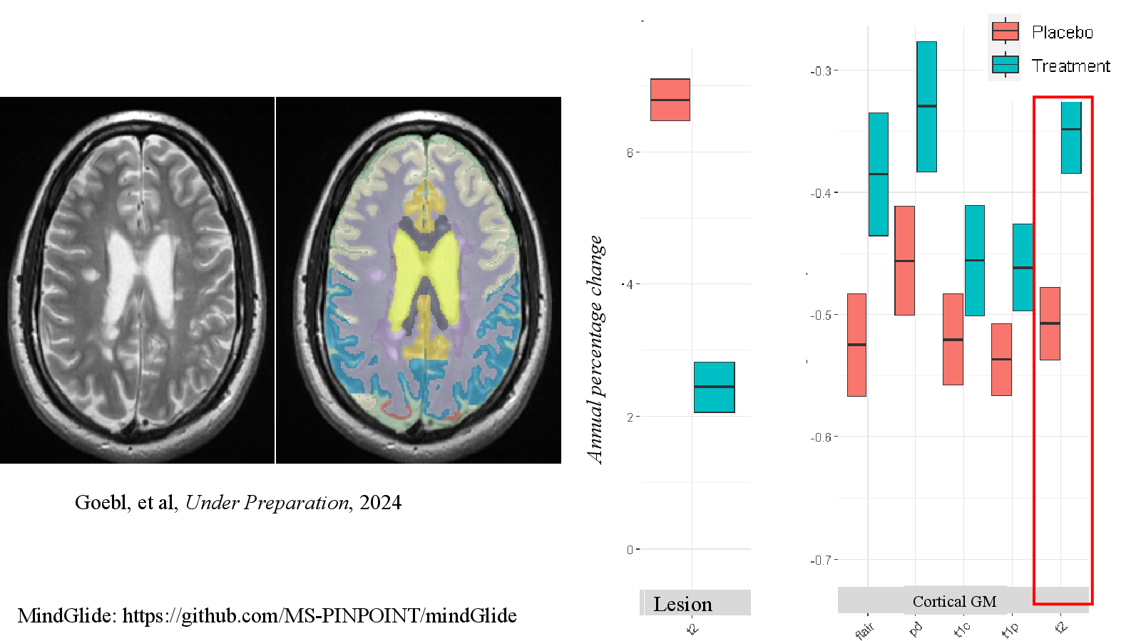
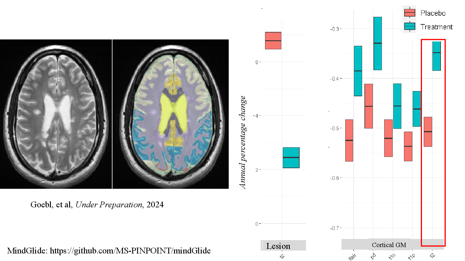
MindGlide: https://github.com/MS-PINPOINT/mindGlide
Slide 08
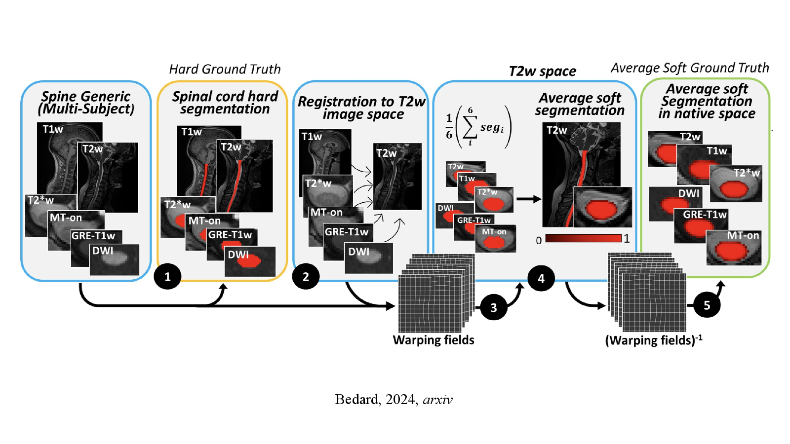
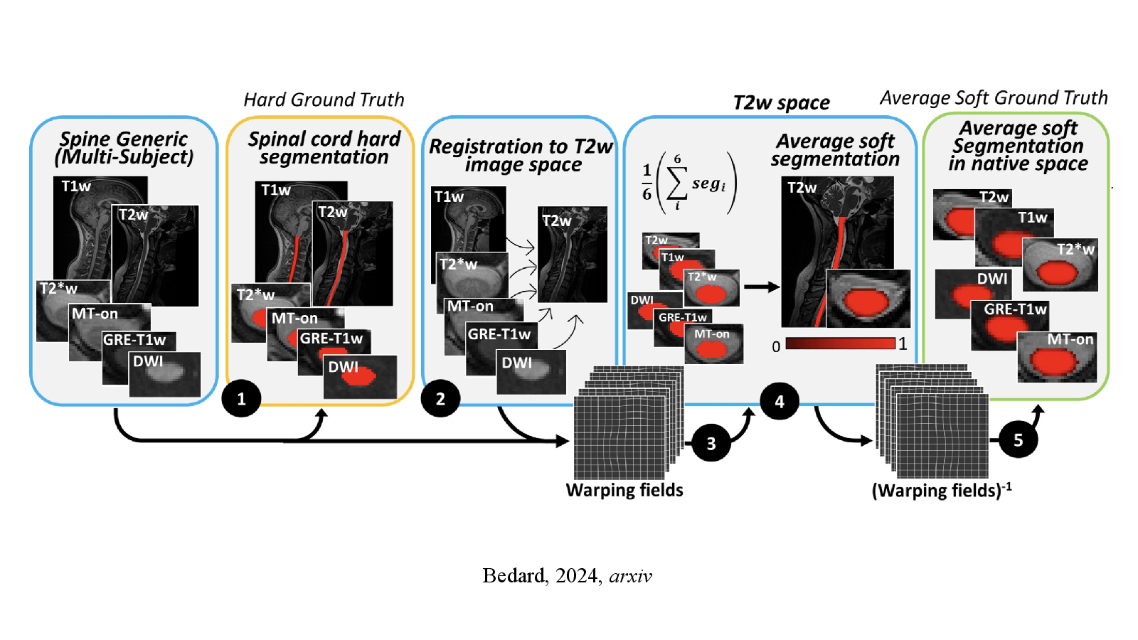
Bedard, 2024, arxiv
Slide 09
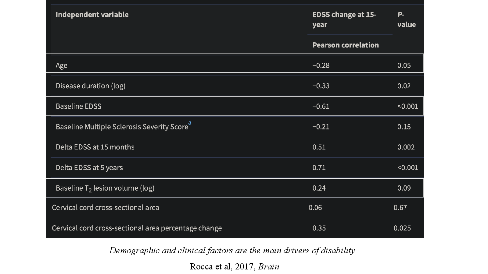
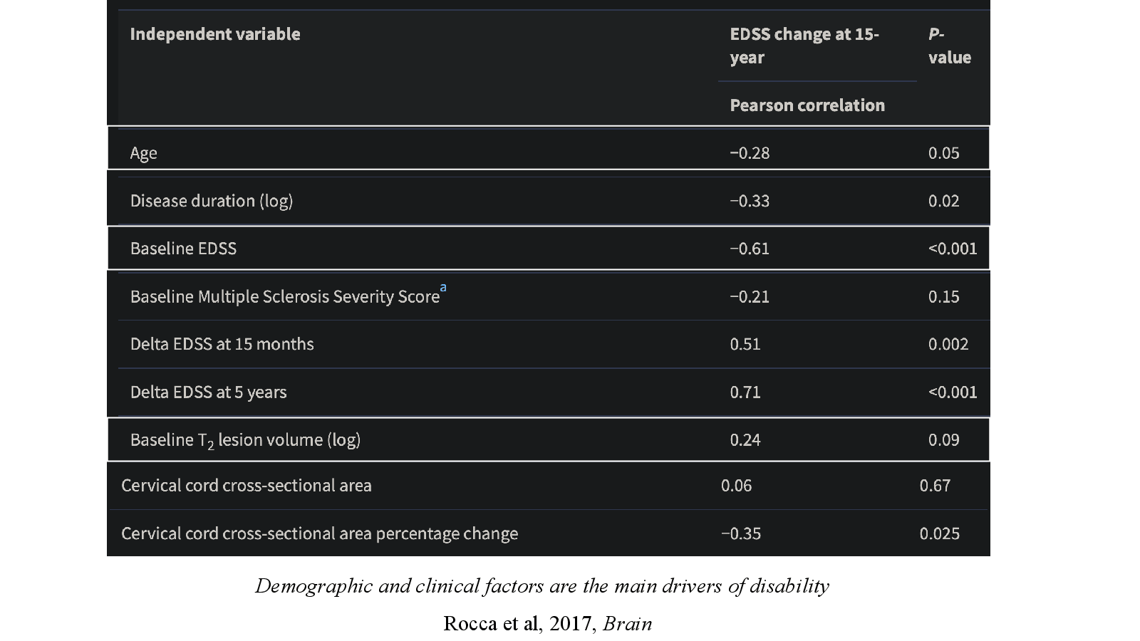
Rocca et al, 2017, Brain
Slide 10
Routine-care MRI for prognosis
Slide 11
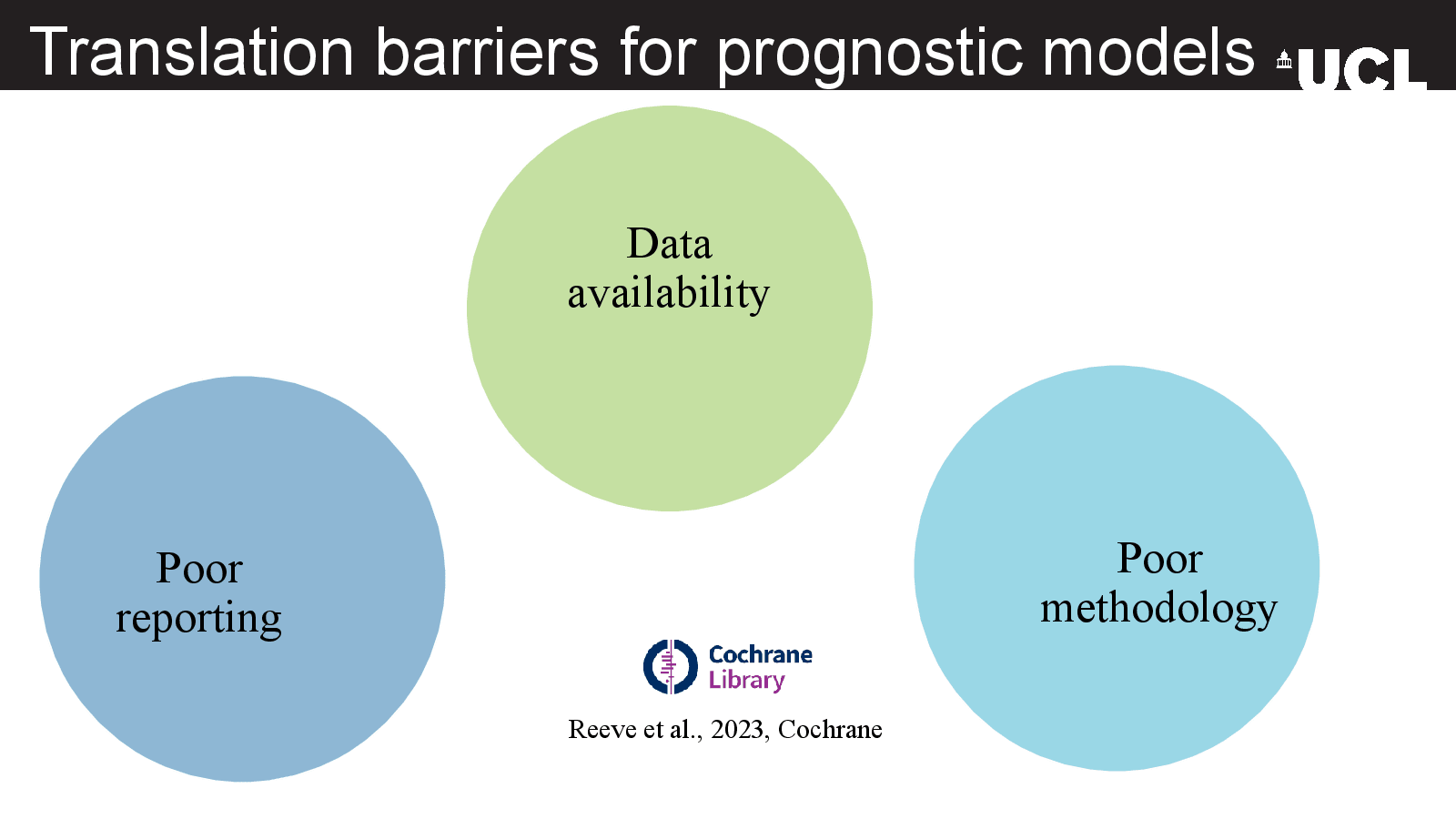
Translation barriers for prognostic models
Slide 12
Next steps
Slide 13
Take home
Slide 14
Slide 14