All talks & lectures

Conference ·

AI Applied to MRI: Promises and Challenges

An ECTRIMS talk on AI across MRI diagnosis, monitoring, prognosis and biological subtyping in multiple sclerosis.

ECTRIMS 2025 · Barcelona, Spain

A web sketch of the deck’s central idea
Delivered
Event
ECTRIMS 2025
Place
Barcelona, Spain
Format
Conference
Source deck
17 slides

From the slides

Deck notes

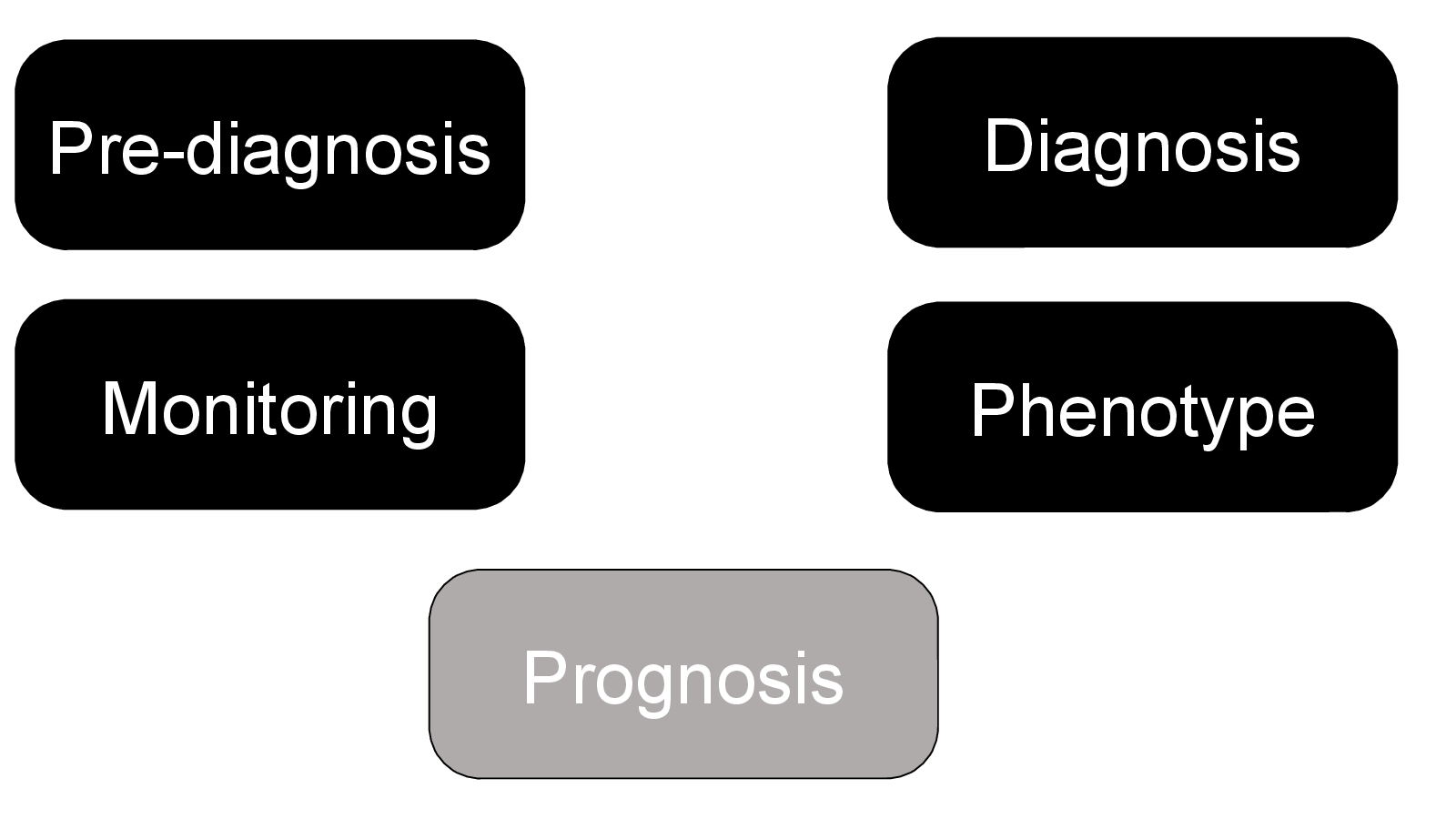
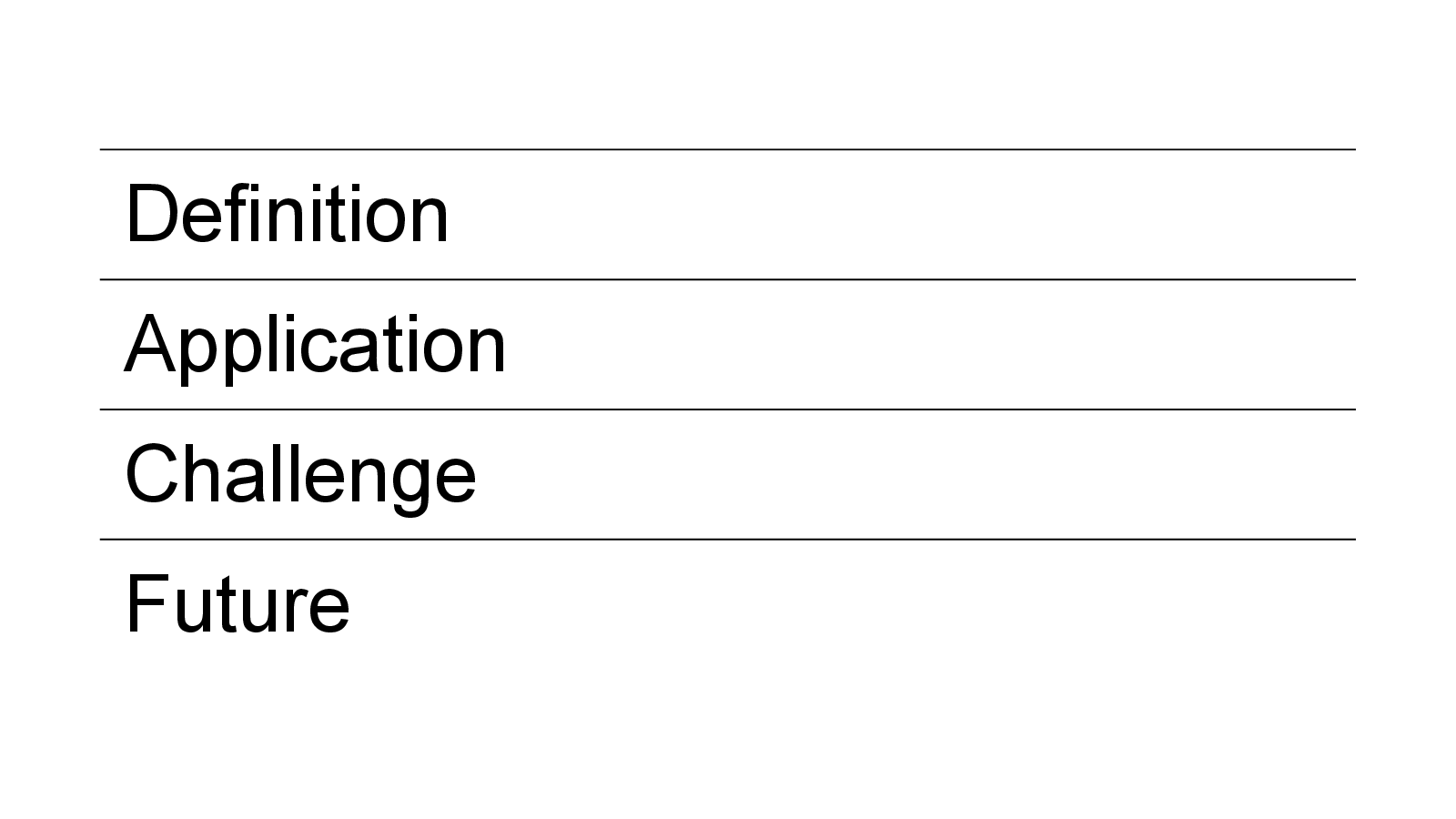
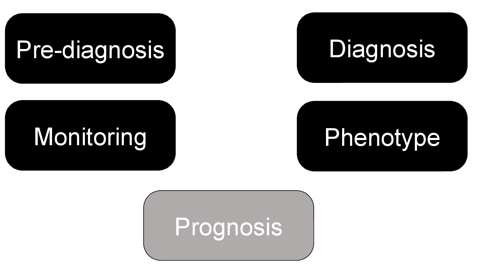
This talk followed AI across the MRI pathway in MS: pre-diagnosis, diagnosis, monitoring, prognosis and phenotype. It compared promising research applications with the narrower set of tools approaching routine clinical use.

The question

Where is AI applied to MRI mature enough to support clinical work, and where does it remain a research method?

Deck outline

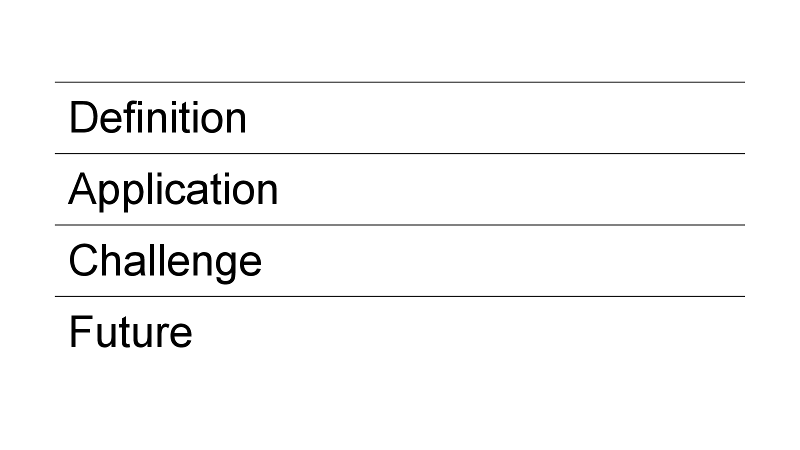
  • Detecting recent lesion activity from conventional MRI
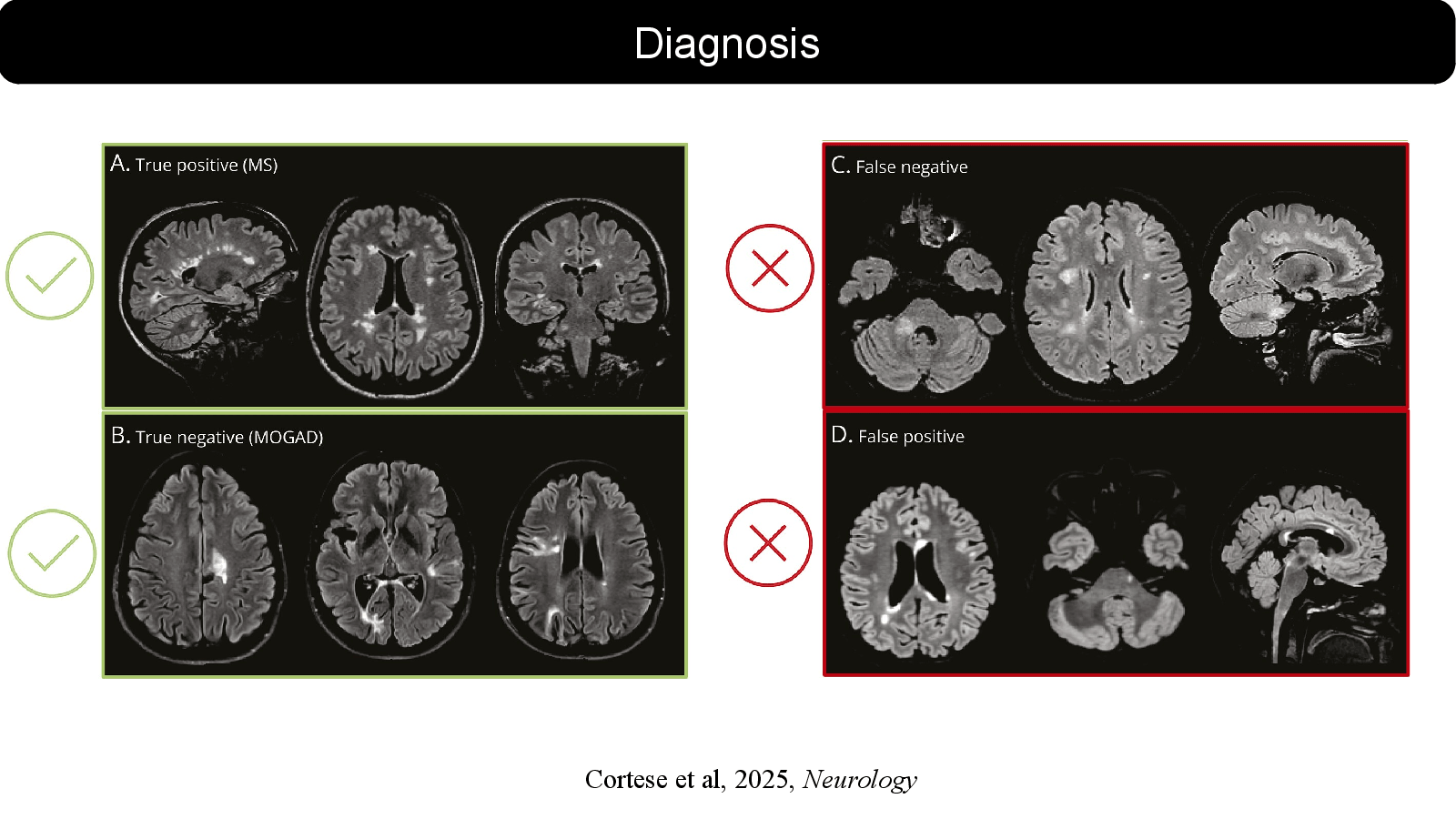
  • Automated support for diagnosis and lesion segmentation
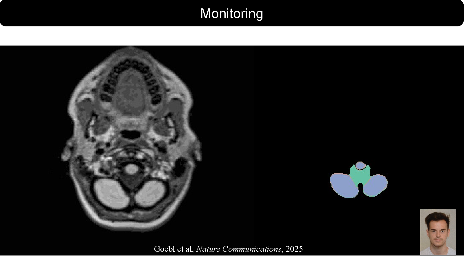
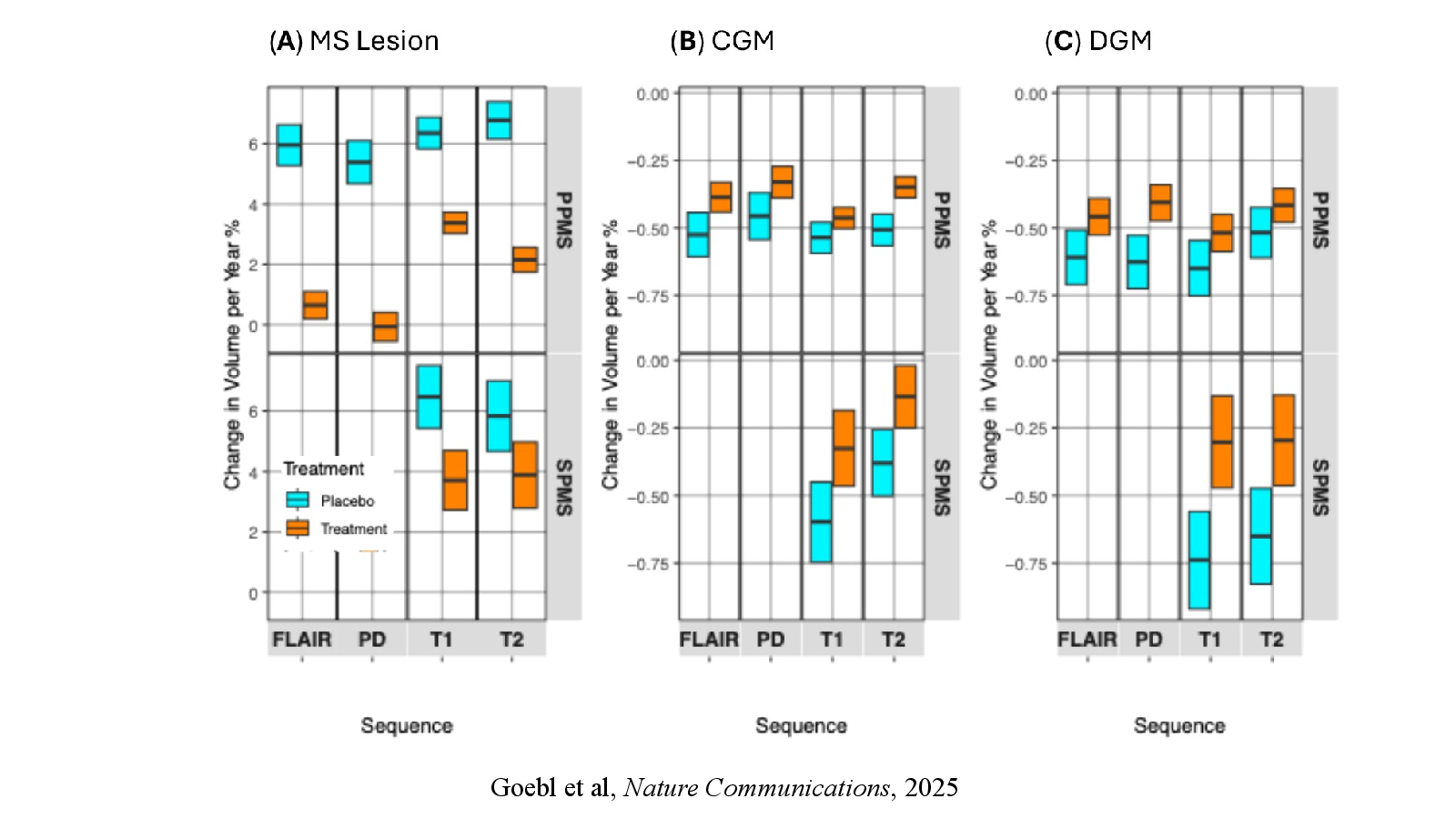
  • Quantitative MRI measures for disease monitoring
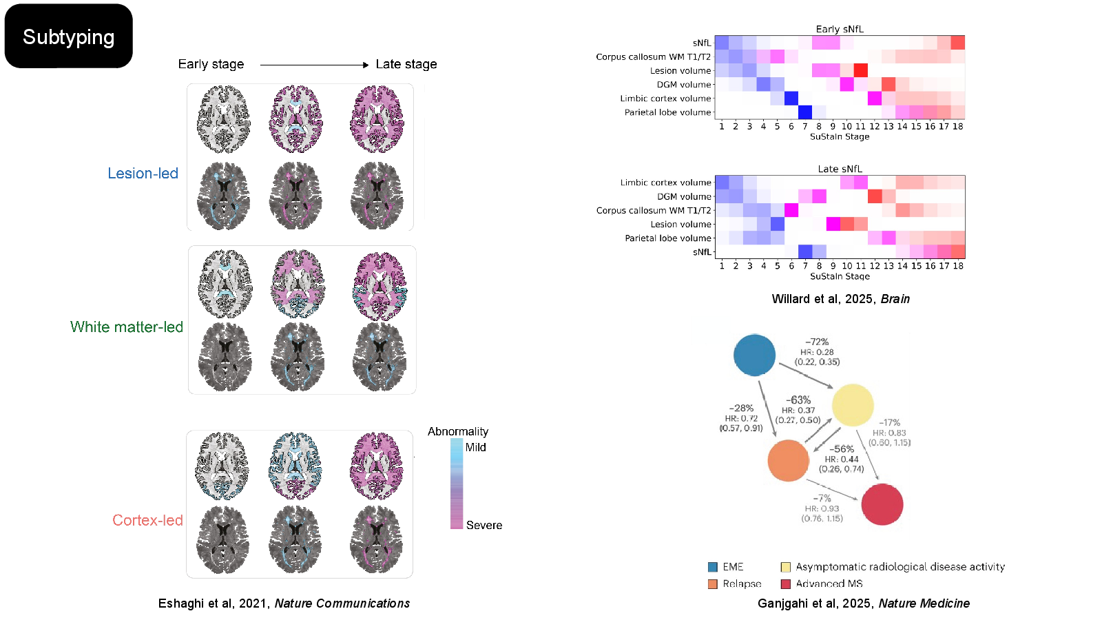
  • Data-driven and multimodal MS subtypes
  • Prognostic models for disease course
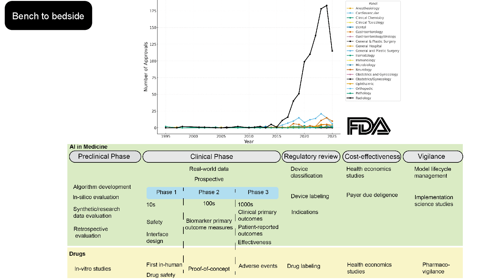
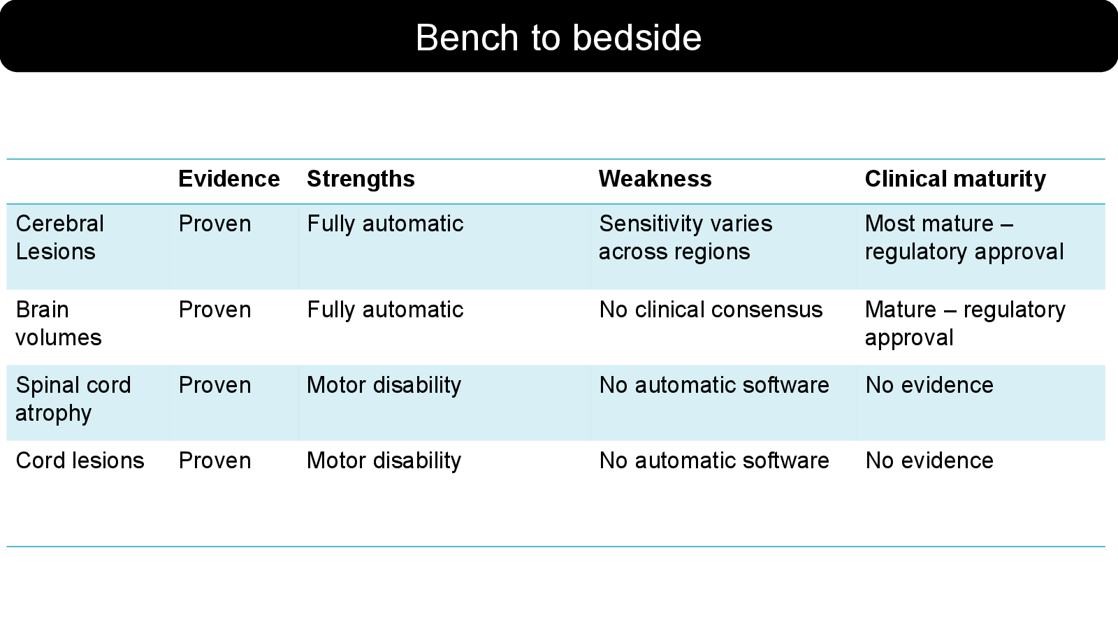
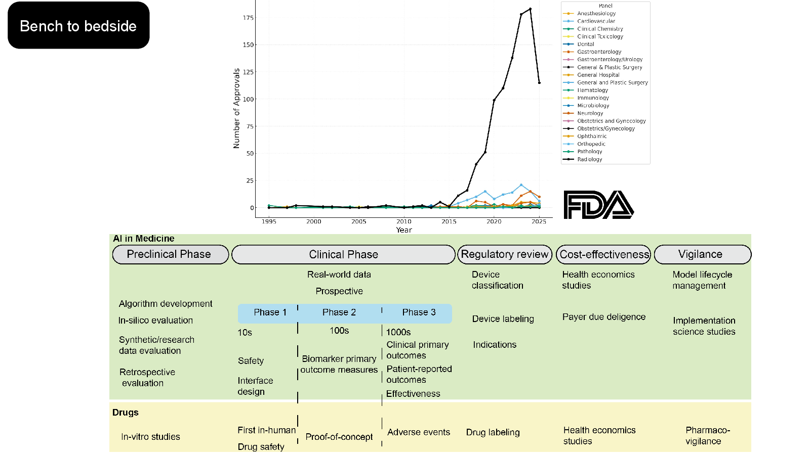
  • Evidence and clinical maturity from bench to bedside

Take-away

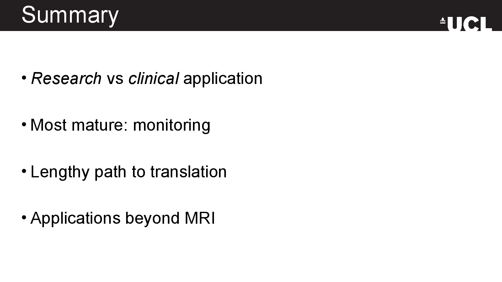
Automated monitoring is the most mature application presented; diagnosis, prognosis and subtyping still face a longer path to clinical translation.

Original deck

Slide by slide

Read the 17-slide presentation in order, or use the index to jump.

  1. Slide 01

    AI applied to MRI

    Slide 1: AI applied to MRIOpen full-size slide

    Promises and challenges

    Arman Eshaghi, MD, PhD

    Queen Square Institute of Neurology

    University College London

    UK

  2. Slide 02

    Conflicts of interest

    Slide 2: Conflicts of interestOpen full-size slide
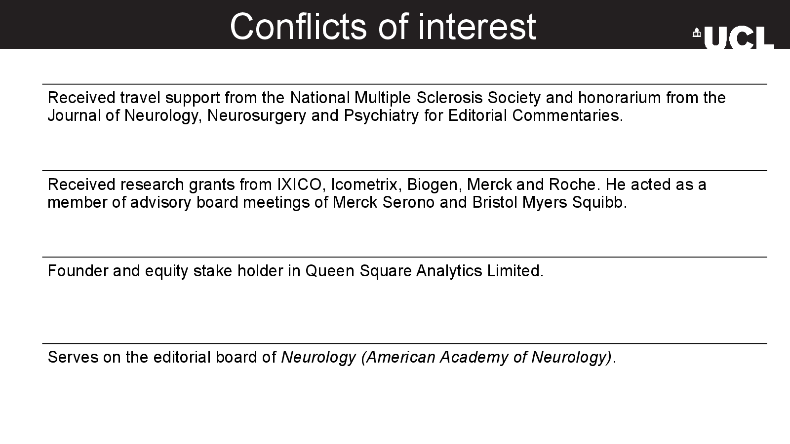
    • Received travel support from the National Multiple Sclerosis Society and honorarium from the Journal of Neurology, Neurosurgery and Psychiatry for Editorial Commentaries.
    • Received research grants from IXICO, Icometrix, Biogen, Merck and Roche. He acted as a member of advisory board meetings of Merck Serono and Bristol Myers Squibb.
    • Founder and equity stake holder in Queen Square Analytics Limited.
    • Serves on the editorial board of Neurology (American Academy of Neurology).
  3. Slide 06

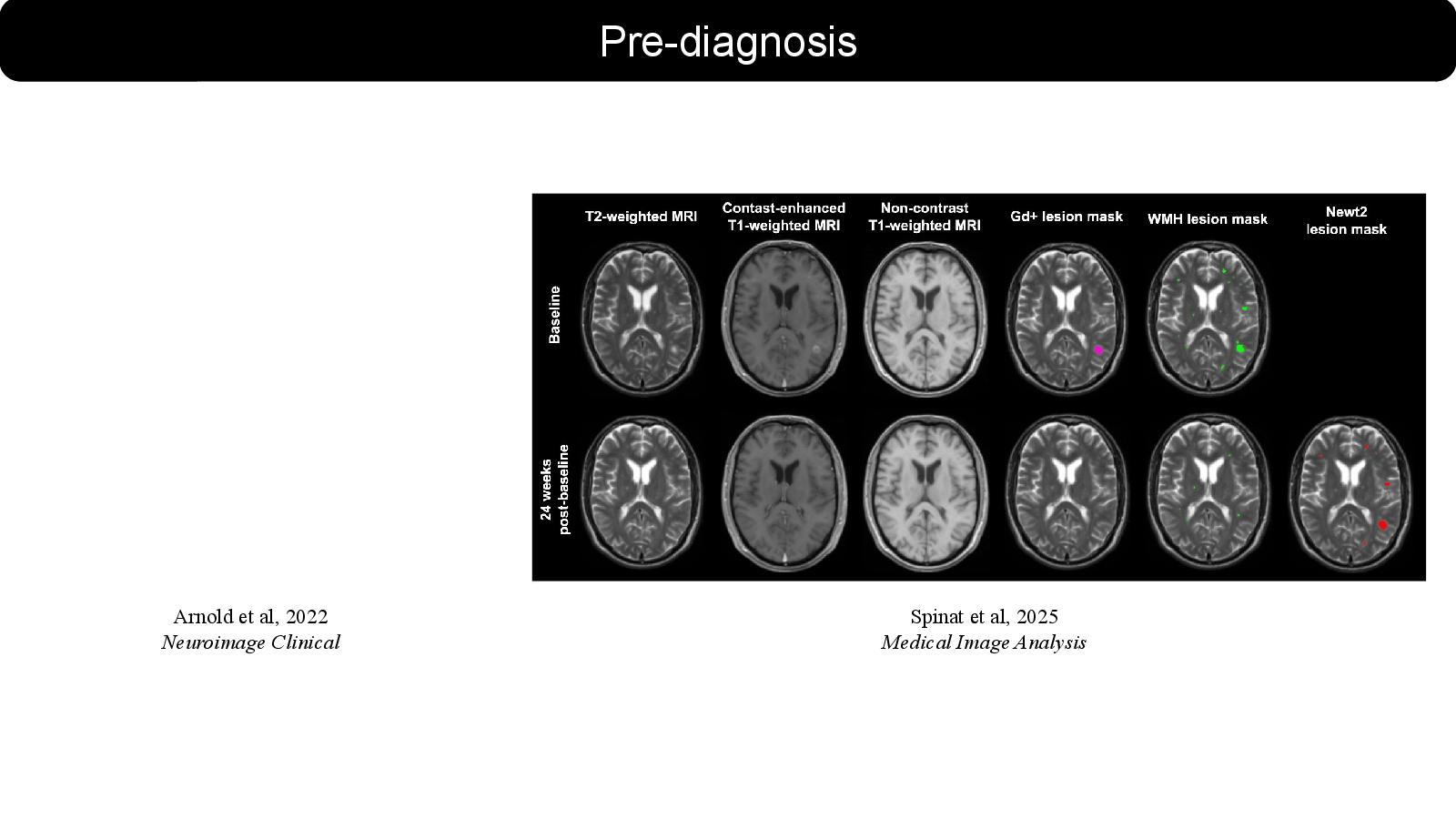
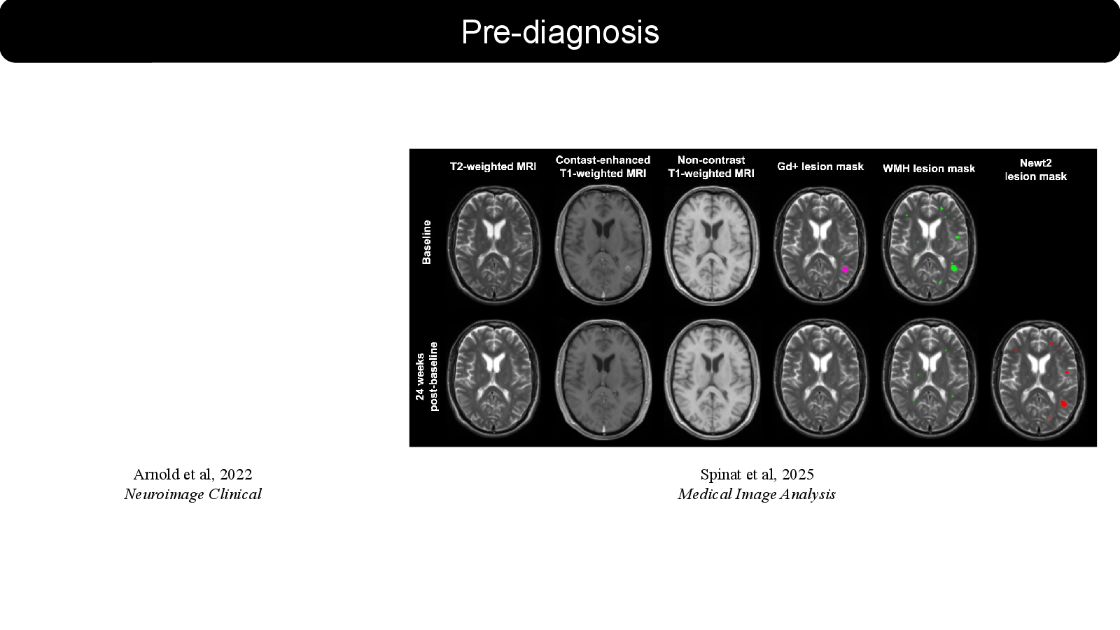
    Arnold et al, 2022

    Slide 6: Arnold et al, 2022Open full-size slide

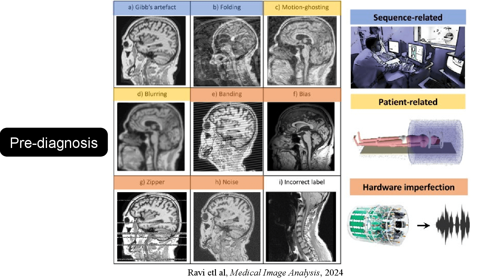
    Pre-diagnosis

    Neuroimage Clinical

    Spinat et al, 2025

    Medical Image Analysis

    Speaker notes
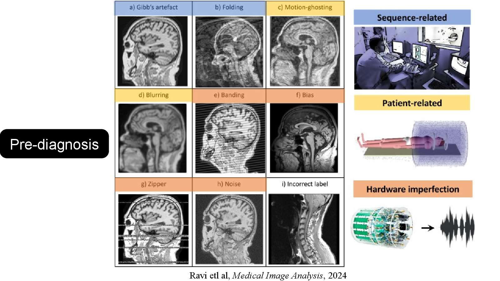
    1. Multiple sclerosis (MS) is a chronic inflammatory disease characterized by demyelinating lesions in the central nervous system. Cross-sectional measurements of acute inflammatory lesion activity are typically obtained by detecting the presence of gadolinium enhancement in lesions, which typically lasts 3-6 weeks. We formulate the novel and clinically relevant task of quantification of recent acute lesion activity from the past 24 weeks (6 months) using single-timepoint conventional brain magnetic resonance imaging (MRI). We develop and compare several deep learning (DL) methods for estimating this brain-level acuteness score and show that a 2D-UNet can accurately predict acute disease activity at the patient-level while outperforming transformers and ensemble approaches. In the context of identifying subjects with acute (less than 6 months-old) lesion activity, our 2D-UNet achieves an area under the receiver-operating curve in the range on independent relapsing-remitting MS cohorts. When used in conjunction with measurements of gadolinium-enhancing lesion activity, our model significantly improves the prognostication of future acute lesion activity (over the next 6 months). This model could thus be leveraged for population recruitment in clinical trials to identify a higher number of patients with acute inflammatory activity than current standard approaches (e.g., gadolinium positivity) with a predictable precision/recall trade-off.

    References

  4. Slide 09

    La Rosa et al, 2022, Neuroimage: Clinical
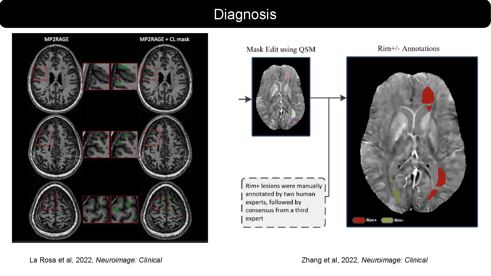
    Slide 9: La Rosa et al, 2022, Neuroimage: ClinicalOpen full-size slide

    Diagnosis

    Zhang et al, 2022, Neuroimage: Clinical

    Speaker notes

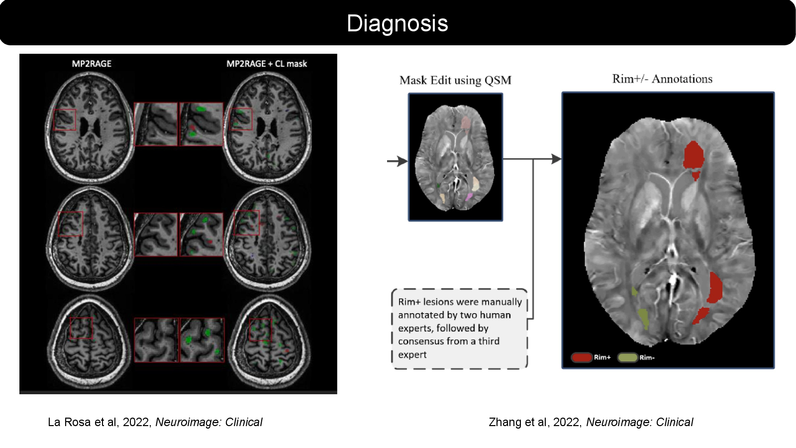
    Three representative axial slices from one MS patient showing the CL segmentation results of an automated CL segmentation method (La Rosa et al., 2020). 7 T MP2RAGE (left column) and CL mask (right column) showing true positives (green), false negatives (red), and false positives (blue) of the automated approach with respect to the CL manual segmentation. (For interpretation of the references to colour in this figure legend, the reader is referred to the web version of this article.)

    References

  5. Slide 13

    Eshaghi et al, 2021, Nature Communications
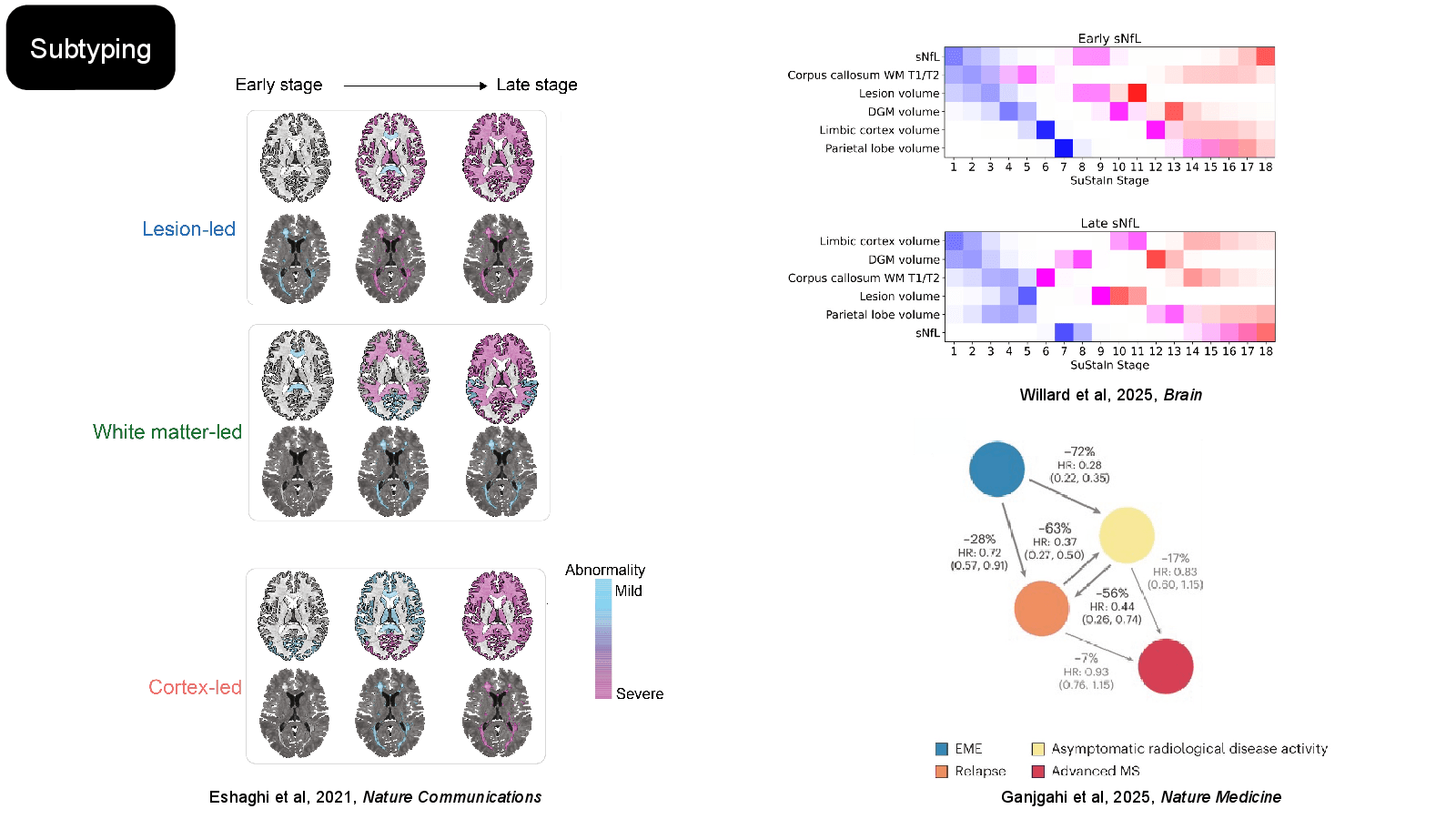
    Slide 13: Eshaghi et al, 2021, Nature CommunicationsOpen full-size slide

    Subtyping

    Willard et al, 2025, Brain

    Ganjgahi et al, 2025, Nature Medicine

    Speaker notes

    Gross et al. employed this method to uncover three groups based on immune cell markers in early MS, with different patterns in CD4, CD8 T cells, natural killer cell compartments, and cytokines[25]. These endotypes showed subtle variations in disease course and treatment response, suggesting potential future clinical applications

    References

  6. Slide 15

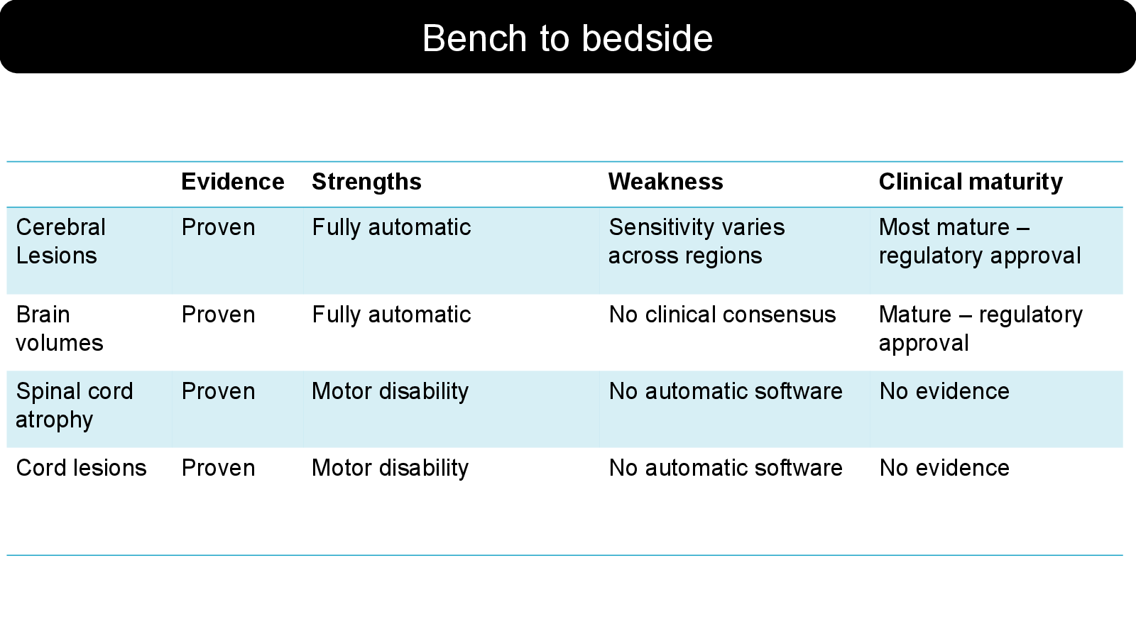
    Bench to bedside

    Slide 15: Bench to bedsideOpen full-size slide
    • Evidence — Strengths — Weakness — Clinical maturity
    • Cerebral Lesions — Proven — Fully automatic — Sensitivity varies across regions — Most mature – regulatory approval
    • Brain volumes — Proven — Fully automatic — No clinical consensus — Mature – regulatory approval
    • Spinal cord atrophy — Proven — Motor disability — No automatic software — No evidence
    • Cord lesions — Proven — Motor disability — No automatic software — No evidence
  7. Slide 16

    Summary

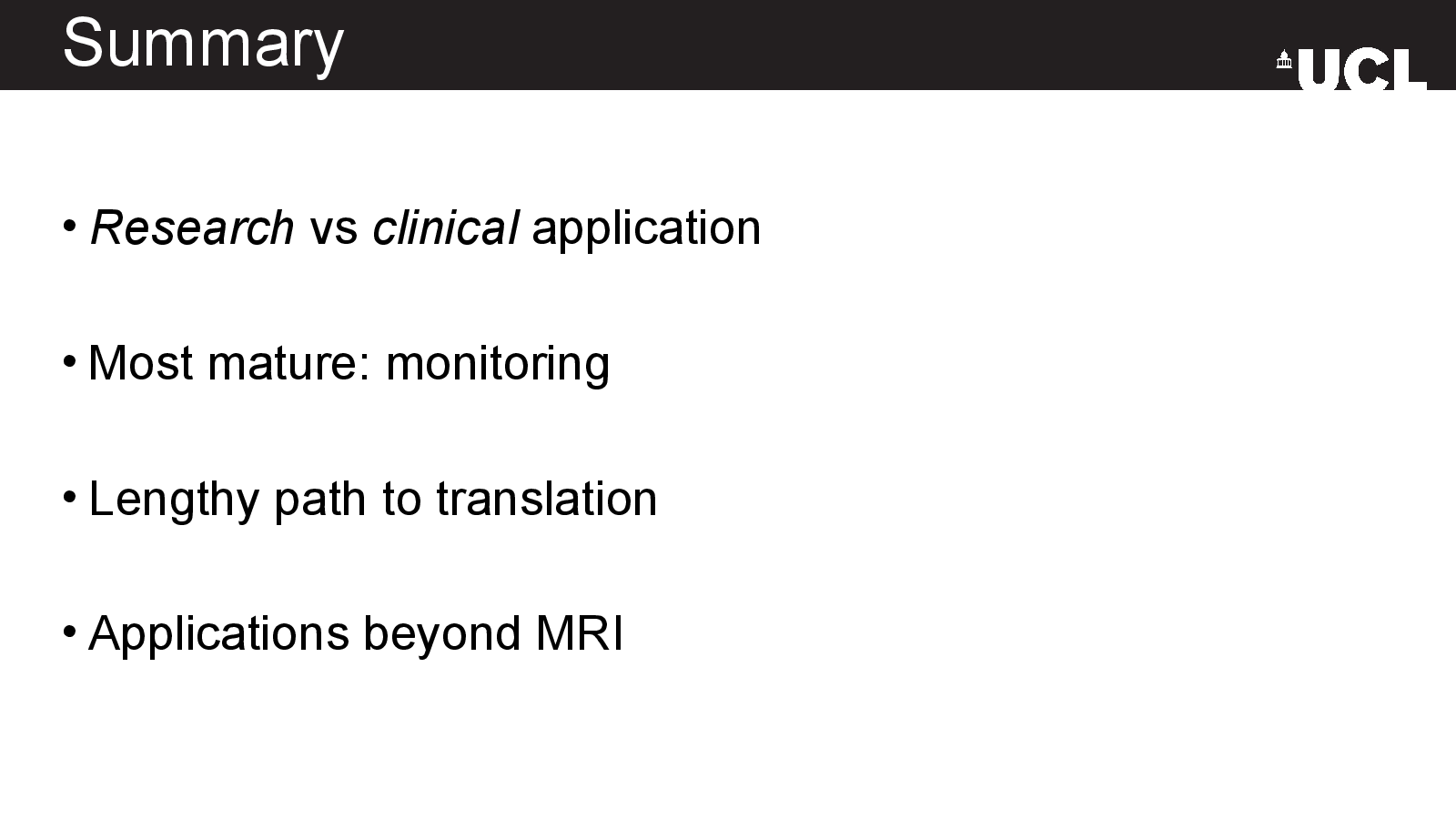
    Slide 16: SummaryOpen full-size slide

    Research vs clinical application

    Most mature: monitoring

    Lengthy path to translation

    Applications beyond MRI